ACCESSORIES1 AND THEIR USES:
EYEPIECES:
 Although most microscopes were supplied with several different
eyepieces, or oculars, specialized eyepieces were also not uncommon.
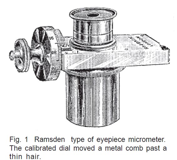
Among these were the eyepiece micrometers. There were two basic types.
Early on, the Ramsden or filar type (Fig. 1), was often used, through which the user could see a
row of metal points resembling a comb. Riding in front of these was a
hair, usually of spider web, which could be moved across the combs via
a calibrated screw and through which objects could then be measured.
Although most microscopes were supplied with several different
eyepieces, or oculars, specialized eyepieces were also not uncommon.
Among these were the eyepiece micrometers. There were two basic types.
Early on, the Ramsden or filar type (Fig. 1), was often used, through which the user could see a
row of metal points resembling a comb. Riding in front of these was a
hair, usually of spider web, which could be moved across the combs via
a calibrated screw and through which objects could then be measured.
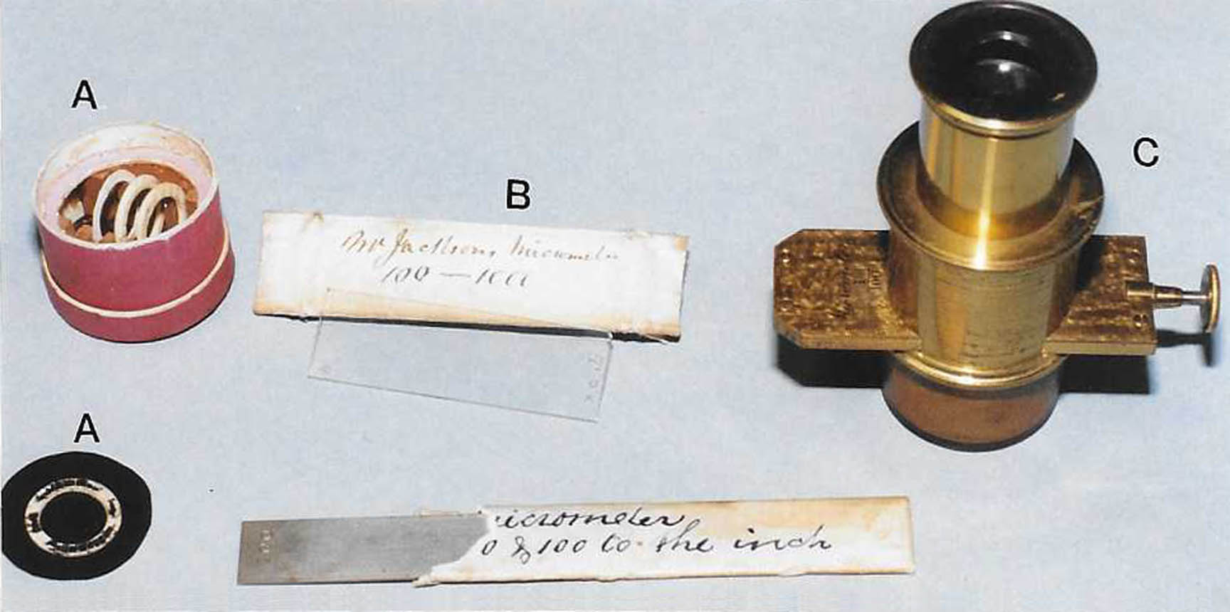 A simpler and much less expensive method was developed later once
accurate dividing
engines could scribe a series of parallel lines on glass. Such glass
pieces were inserted through a slit in the eyepiece (Fig. 2C) and used
directly, much the same way as our modern eyepiece micrometers are today. The type which fit through the slit in the eyepiece became known as a Jackson Micrometer.
Another variant was a round drop-in micrometer which could be
placed inside the eyepiece after unscrewing part of it.
A simpler and much less expensive method was developed later once
accurate dividing
engines could scribe a series of parallel lines on glass. Such glass
pieces were inserted through a slit in the eyepiece (Fig. 2C) and used
directly, much the same way as our modern eyepiece micrometers are today. The type which fit through the slit in the eyepiece became known as a Jackson Micrometer.
Another variant was a round drop-in micrometer which could be
placed inside the eyepiece after unscrewing part of it.
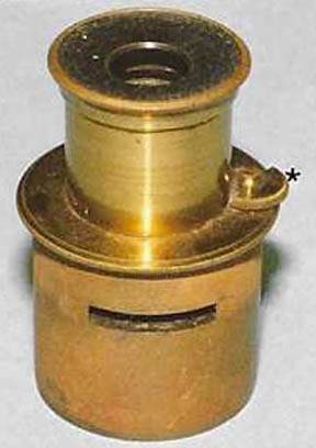 Another accessory that seems inappropriately uncommon is the use of a
pointer inside the eyepiece (Fig. 3). These can be recognized by the
little piece of metal rotating on a screw on the outside of the
eyepiece with the screw’s axis parallel to the optical path. Some other
variations include a fixed pointer, and others.
Another accessory that seems inappropriately uncommon is the use of a
pointer inside the eyepiece (Fig. 3). These can be recognized by the
little piece of metal rotating on a screw on the outside of the
eyepiece with the screw’s axis parallel to the optical path. Some other
variations include a fixed pointer, and others.
 In addition, several ingenious makers devised attachments to fit on or
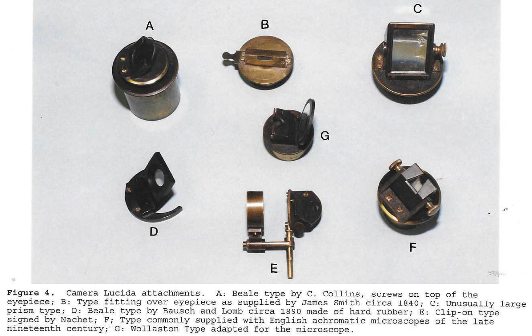
over their oculars. A typical example was the camera lucida, several examples of which are shown in figure 4. These are drawing aids which allow one to see their subject and the drawing they are making at the same time; this results in more accurate imaging. For more examples and more information please see the dedicated web page article about camera lucidas for the microscope.
In addition, several ingenious makers devised attachments to fit on or
over their oculars. A typical example was the camera lucida, several examples of which are shown in figure 4. These are drawing aids which allow one to see their subject and the drawing they are making at the same time; this results in more accurate imaging. For more examples and more information please see the dedicated web page article about camera lucidas for the microscope.
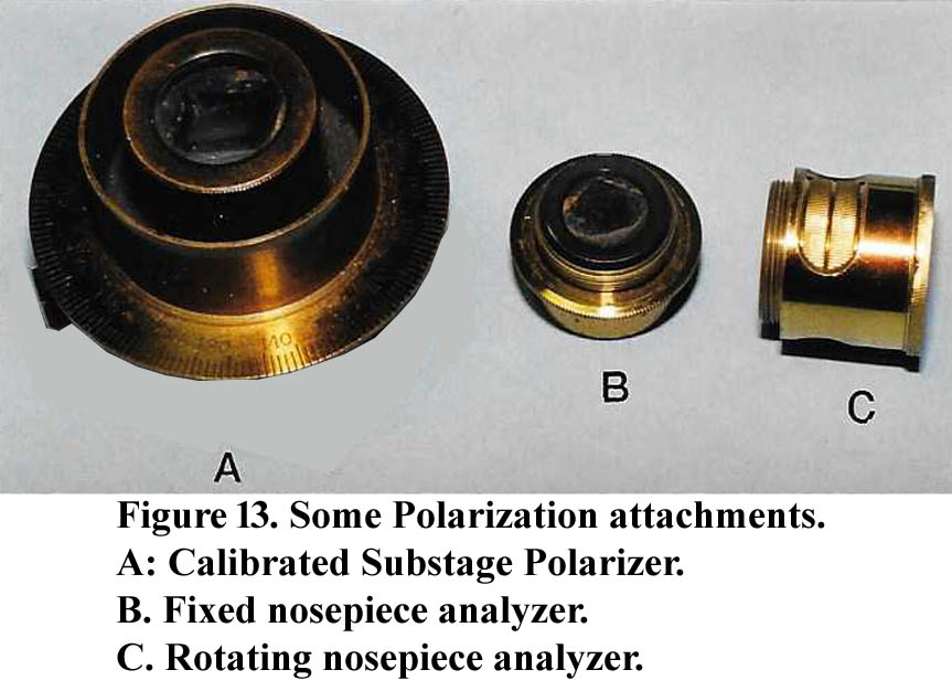
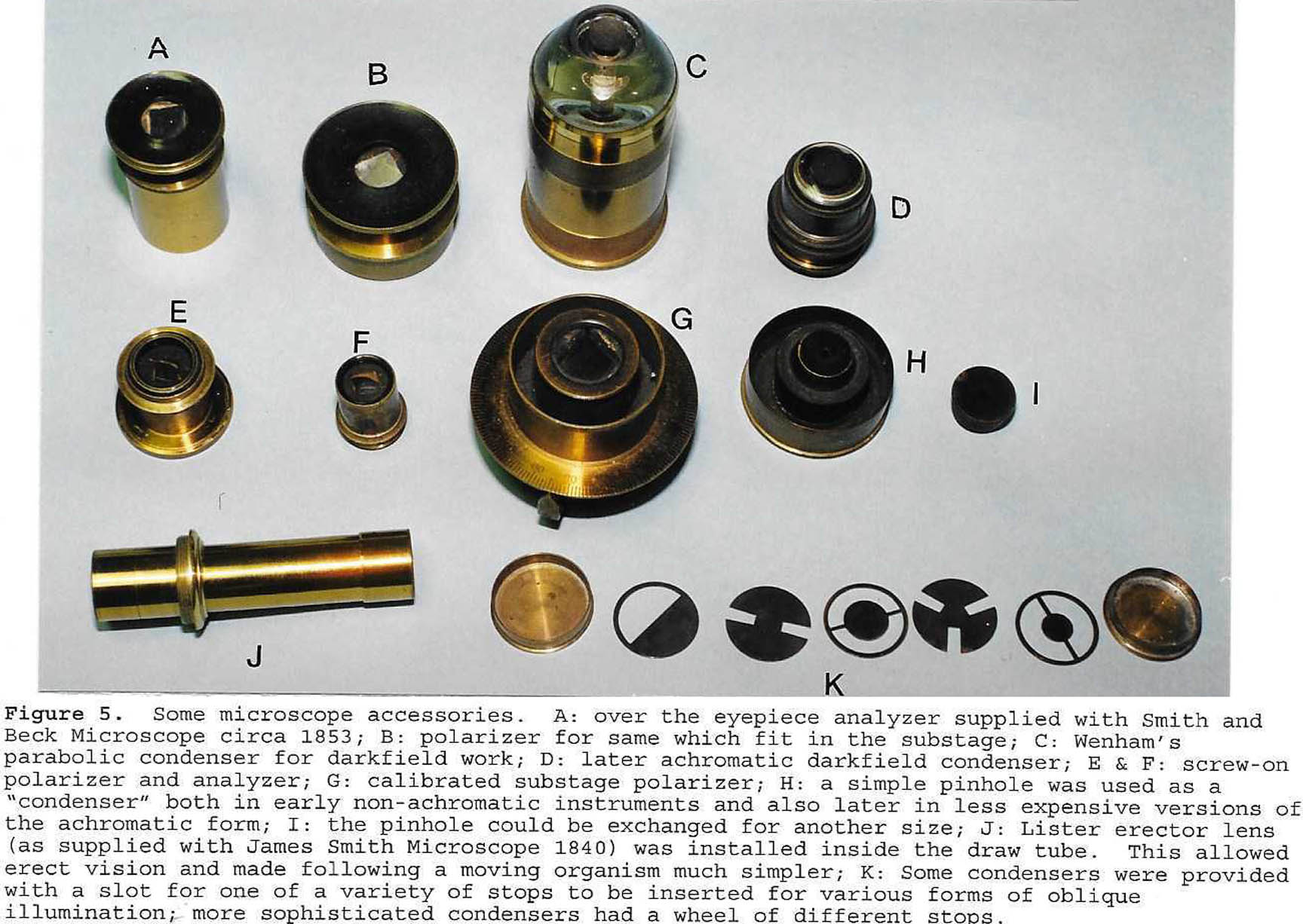 Another over the eyepiece attachment was the analyzer (Fig. 5) for polarized light microscopy. Although sometimes the analyzer sat above the ocular, it was most commonly made as a replacement for the cap on the ocular(Fig 5A). Occasionally it was also made in a form that could be attached between the optical tube and the objective with an adapter which allowed it to rotate(Fig 13C).
It could also take the form of a slider which fit through a slit above the nosepiece, and this is a common method even today. The polarizer usually fit into the substage (Figs. 5B, 5G). Related accessories included the double image prism for demonstrating the principals of birefringence, and 'selenites' which are thin plates of the mineral Selenite used as wave retarders. Used with crossed polars, the selenites would impart colors to an anisotropic subject. Selenites were provided as a single retarder, built in to slide holder(a 'selenite stage'), or in a device which could combine the selenites and rotate them such as in the Darker's Selenite Stage, or a substage stack of rotating selenites.
Another over the eyepiece attachment was the analyzer (Fig. 5) for polarized light microscopy. Although sometimes the analyzer sat above the ocular, it was most commonly made as a replacement for the cap on the ocular(Fig 5A). Occasionally it was also made in a form that could be attached between the optical tube and the objective with an adapter which allowed it to rotate(Fig 13C).
It could also take the form of a slider which fit through a slit above the nosepiece, and this is a common method even today. The polarizer usually fit into the substage (Figs. 5B, 5G). Related accessories included the double image prism for demonstrating the principals of birefringence, and 'selenites' which are thin plates of the mineral Selenite used as wave retarders. Used with crossed polars, the selenites would impart colors to an anisotropic subject. Selenites were provided as a single retarder, built in to slide holder(a 'selenite stage'), or in a device which could combine the selenites and rotate them such as in the Darker's Selenite Stage, or a substage stack of rotating selenites.
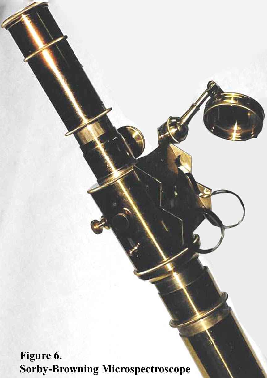
Finally, there were some very sophisticated microspectroscopes
which were first devised by Sorby and constructed by John Browning
(Fig. 6). These had varying degrees of complexity, and could be fitted
with a micrometer for precise measurements of the spectra. They usually
had some provision for comparison to a standard, often a liquid in a
glass tube held onto the side of microspectroscope by spring clips.
THE NOSE
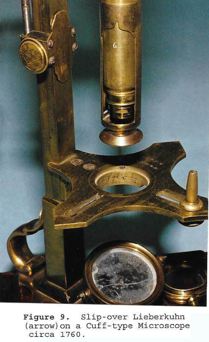
Not only were objectives (Fig. 32) of varying focal length usually supplied, but some were either more specialized, or had provision for further attachments. Perhaps the earliest specialized objective was the objective with a built-in Lieberkuhn. Although initially intrinsic to objectives, Cuff soon
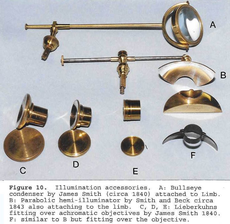
developed the slip-over Lieberkuhn reflector that could be put on or removed; these were supplied with early nonachromatic instruments such as the ‘Cuff Double Microscope’ (Fig. 9). Later slip-over Lieberkuhns were supplied for achromatic objectives (Fig. 10 C,D & E).
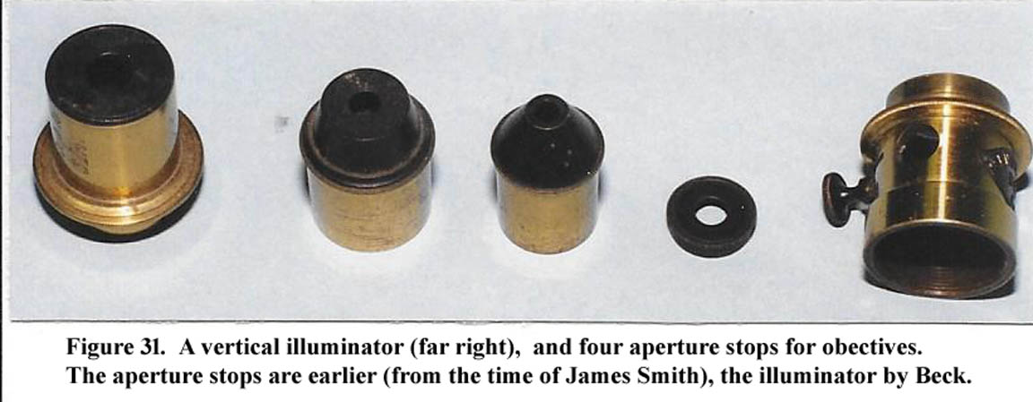
Lieberkuhns, however, were not ideal illuminators for
opaque objects , and various types of vertical illuminators were developed. An example by Richard Beck is shown in Figure 31. It used a thin circular disc of glass
to allow light to enter and be reflected downward
through the objective. The user would view the illuminated
object while looking through this same glass.
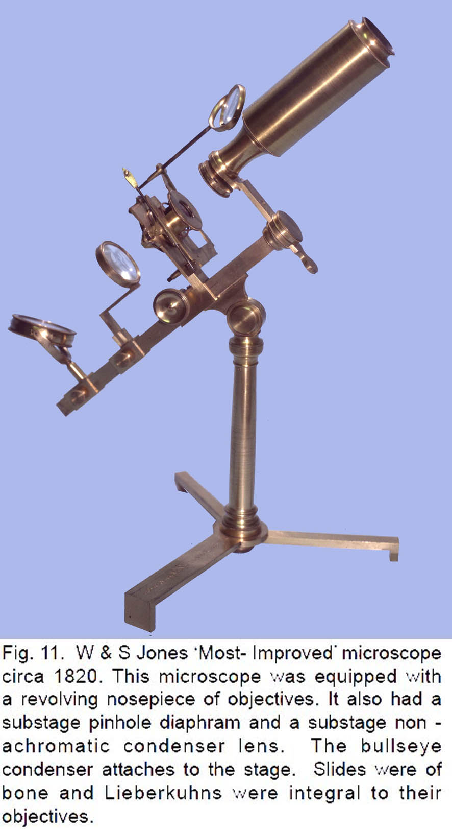
 Although developed quite early, and found on microscopes
by George Adams (1746)3,
Benjamin Martin and W & S Jones (Fig. 11), the use of a multiple
objective nosepiece was not initially
supplied with the achromatic microscopes with
their larger, heavier, and more complex objectives. In
the second half of the nineteenth century, multi-objective
nosepiece attachments were again introduced,
this time accepting the individual objectives (Fig. 12). At first these held two objectives, but later this number gradually increased; in the twentieth century rotating nosepieces could hold as many as six different objectives.
Although developed quite early, and found on microscopes
by George Adams (1746)3,
Benjamin Martin and W & S Jones (Fig. 11), the use of a multiple
objective nosepiece was not initially
supplied with the achromatic microscopes with
their larger, heavier, and more complex objectives. In
the second half of the nineteenth century, multi-objective
nosepiece attachments were again introduced,
this time accepting the individual objectives (Fig. 12). At first these held two objectives, but later this number gradually increased; in the twentieth century rotating nosepieces could hold as many as six different objectives.
Another, but much later nosepiece innovation, was the centerable nosepiece, required for advanced work with polarized light. In this case objectives were stored on individual centerable fittings, each of which was exchanged on the nose as required; exchanging these was made easier by quick-change fitting. Much later, in the twentieth century, a rotating nosepiece became available in which each objective on
it could be centered individually, retaining the advantages of centration, while at the same the convenience of a rotating objective changer.
As noted above, in addition to these attachments, an
accessory was also sometimes supplied which joined
between the nosepiece and the objective and accepted
the analyzer providing an alternative location to the
over-the-eyepiece configuration (Fig. 13C).
INSIDE THE TUBE
Although many microscopists and collectors are familiar with the
field lens mounted inside the main ocular tube to give
a wider field of view, few are familiar with another
attachment called the Lister erecting lens system, or
simply the erector lens. This narrow optical arrangement
(Fig. 5J) is often attached to the draw tube, screwing
into its bottom. This accessory allowed easier
manipulation of the stage, when following a moving
organism, since movements were in the same apparent
direction as that of the organism.
SPECIAL SLIDES AND OTHER STAGE FITTINGS
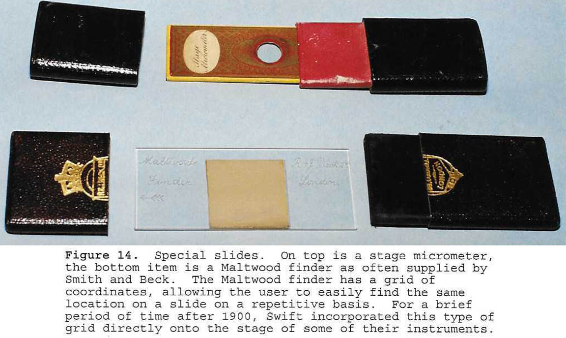 In place of ordinary slides, a variety of measuring devices
were supplied; the more common of which was
the slide micrometer (Fig. 14).
In place of ordinary slides, a variety of measuring devices
were supplied; the more common of which was
the slide micrometer (Fig. 14).
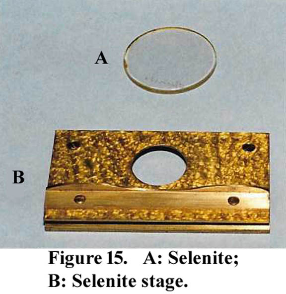 Selenites, used to enhance polarization microscopy,
were supplied either as a slide or in specialized brass
fittings (Fig. 15). Some of these had provision for mechanical
rotation of the selenite or even multiple selenites.
Selenites, used to enhance polarization microscopy,
were supplied either as a slide or in specialized brass
fittings (Fig. 15). Some of these had provision for mechanical
rotation of the selenite or even multiple selenites.
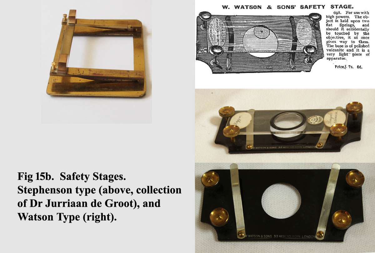
Another type of stage accessory was the safety stage (Fig 15b). These were provided to prevent damage to both slide and objective during overzealous focusing.
Another specialized slide was the finder
. This device, registered on a fixed corner rest on the stage, has a grid, which under the microscope could be used to reposition the stage to a specific point on a slide previously noted at that location on the grid. The most common type was Maltwood's Finder(Fig.14, bottom).
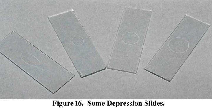
 As for observations of living organisms, a variety of
methods were used. For small organisms, various
shapes and sizes of depression slides were eventually
supplied(Fig. 16). But for greater capacity, various types
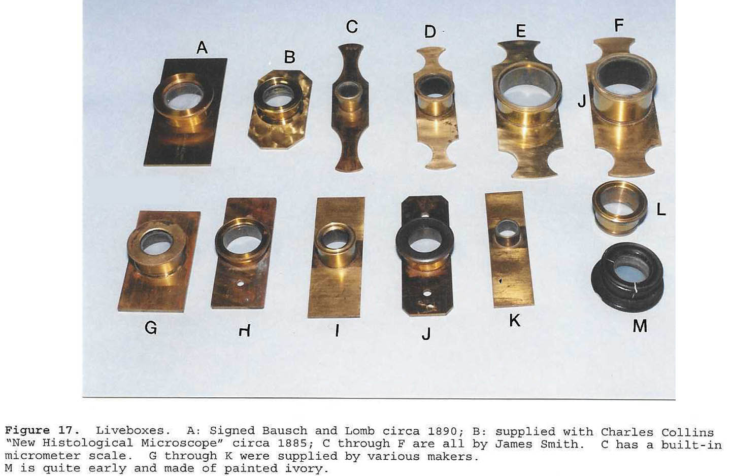
of 'live boxes' or larger glass chambers were supplied.
Figure 17 shows a variety of live boxes that could be
used for wet or dry animals. Early models were constructed
of ivory or brass and would often fit into the
hole in the stage. Later, they were fitted onto a rectangular
brass slide above the stage; rarely, they contained micrometer
markings. An original article on liveboxes is now available on this site.
As for observations of living organisms, a variety of
methods were used. For small organisms, various
shapes and sizes of depression slides were eventually
supplied(Fig. 16). But for greater capacity, various types
of 'live boxes' or larger glass chambers were supplied.
Figure 17 shows a variety of live boxes that could be
used for wet or dry animals. Early models were constructed
of ivory or brass and would often fit into the
hole in the stage. Later, they were fitted onto a rectangular
brass slide above the stage; rarely, they contained micrometer
markings. An original article on liveboxes is now available on this site.
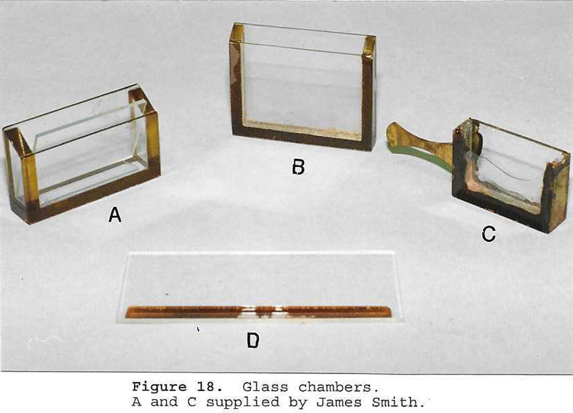
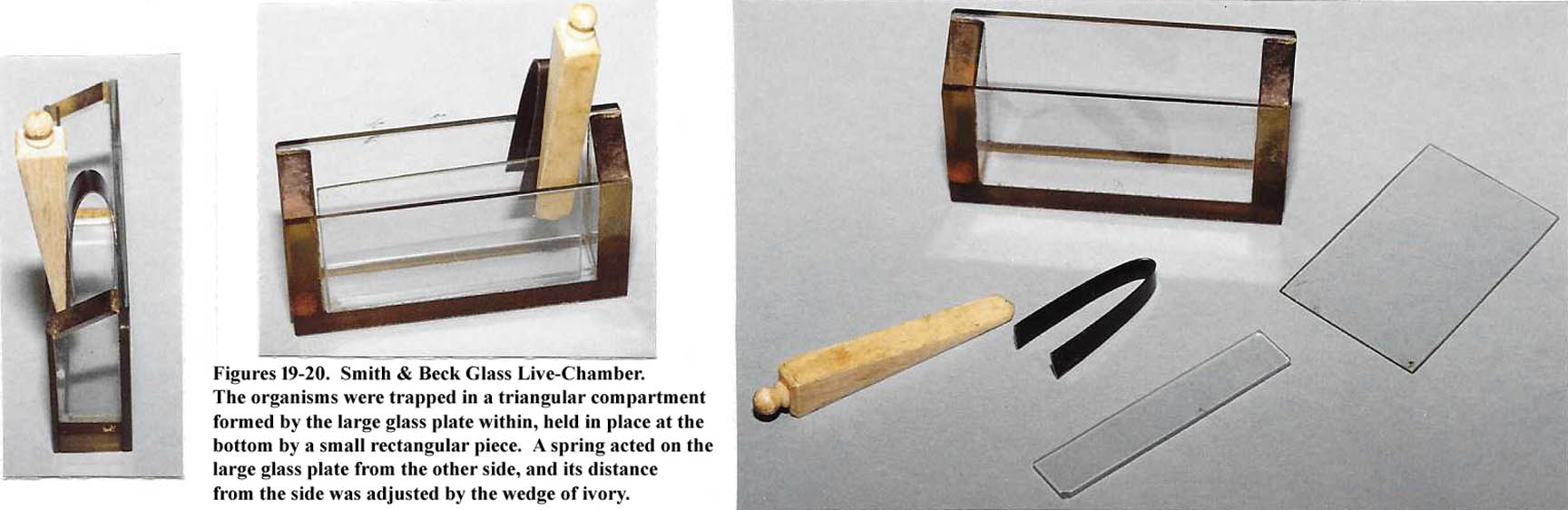 Glass Troughs were supplied by many makers in several
different configurations. Smith supplied one with
a little brass handle (Fig. 18C) and Smith also started a
tradition of supplying a rectangular glass chamber with
an ivory wedge to adjust the space in which the specimen
was trapped (Figs. 19,20).
Glass Troughs were supplied by many makers in several
different configurations. Smith supplied one with
a little brass handle (Fig. 18C) and Smith also started a
tradition of supplying a rectangular glass chamber with
an ivory wedge to adjust the space in which the specimen
was trapped (Figs. 19,20).
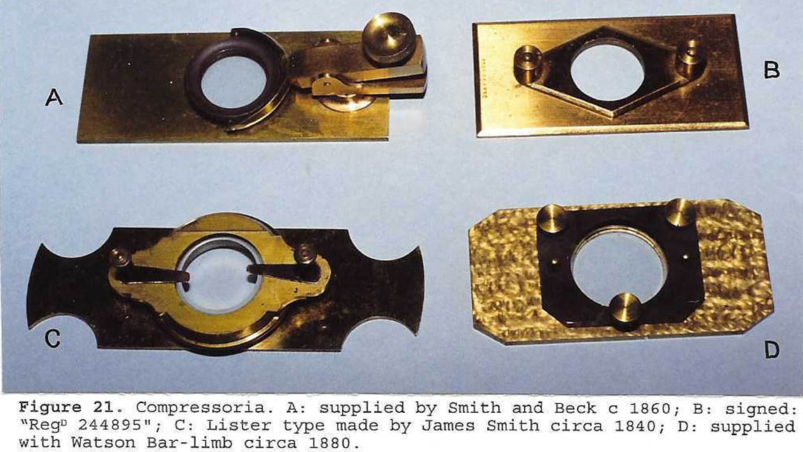 Some living organisms were so active that it was more convenient
to partially immobilize them. This could be accomplished
with a compressor, otherwise known as a 'compressorium,' various types of which are shown in figure 21. For a more comprehensive look at most of the major types of compressoria see the article about compressors added to the site in 2017.
Some living organisms were so active that it was more convenient
to partially immobilize them. This could be accomplished
with a compressor, otherwise known as a 'compressorium,' various types of which are shown in figure 21. For a more comprehensive look at most of the major types of compressoria see the article about compressors added to the site in 2017.
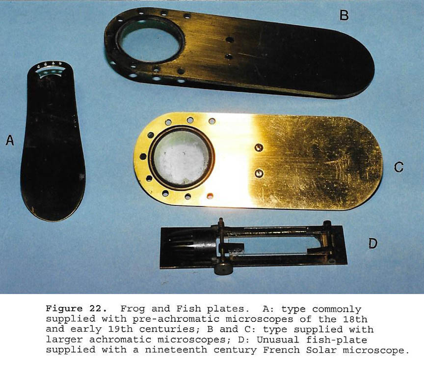
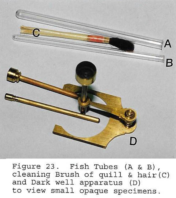 Following the description of the circulation of the
blood by Malpighi, it became popular to view capillary
circulation. This could be seen in the tail of a fish
using a fish-plate(Fig. 22A,22D), in the web of a frog
foot using a frog plate (Fig. 22B, 22C), or in a smaller
fish, tadpole or eel, using a thin test tube (Fig. 23). Please see the dedicated web page article about frogplates for more information.
Following the description of the circulation of the
blood by Malpighi, it became popular to view capillary
circulation. This could be seen in the tail of a fish
using a fish-plate(Fig. 22A,22D), in the web of a frog
foot using a frog plate (Fig. 22B, 22C), or in a smaller
fish, tadpole or eel, using a thin test tube (Fig. 23). Please see the dedicated web page article about frogplates for more information.
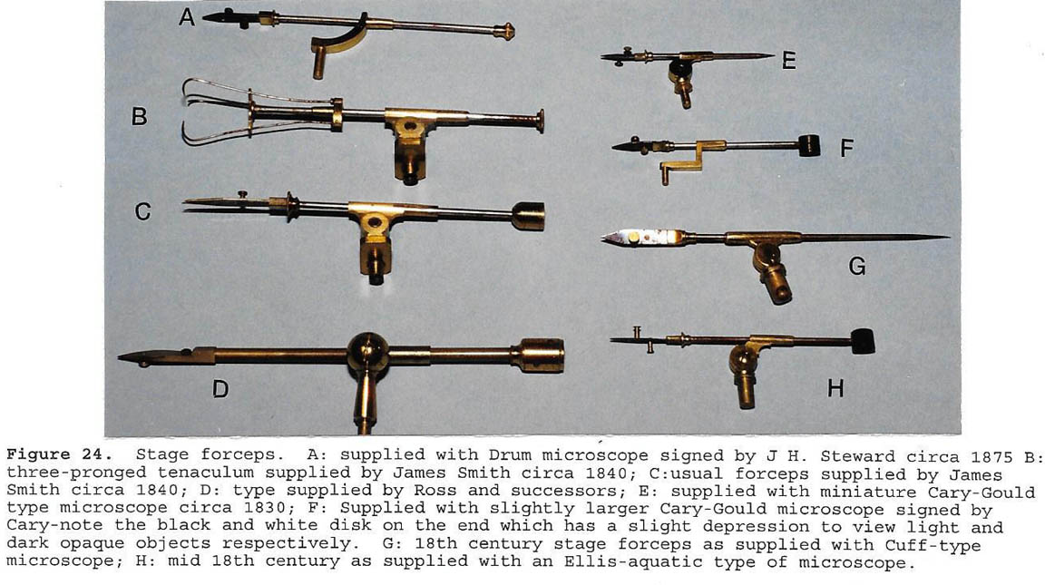 For cruder work, such as looking at a plant or a dead
fly, a stage forceps was often used (Fig. 24). These were most often be attached to a hole on the stage or to the limb assembly. Sometimes they attached to a mechanical stage, or a small rod or other fitting attached to the stage.
For cruder work, such as looking at a plant or a dead
fly, a stage forceps was often used (Fig. 24). These were most often be attached to a hole on the stage or to the limb assembly. Sometimes they attached to a mechanical stage, or a small rod or other fitting attached to the stage.
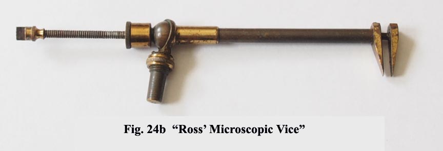
Another variation of the stage forceps, the 'Microscopic Vise'(Fig 24b, collection of Dr Jurriaan de Groot), provides a stronger grasp of the specimen not dependent on a spring, as might be needed for small hard specimens, like a small mineral. Such specimens would likely slip out from between the leaves of the usual stage forceps. This instrument was first reported in the Monthly Microscopical Journal of 1869.
To illuminate opaque objects on the stage, in addition to
Lieberkuhns, the Ross invention of a silver side reflector, or direct illumination by the bullseye condenser were used.
The bullseye condenser could be attached to the stage (Fig. 11), the limb (Fig. 10A), or as a separate accessory on its own
weighted stand (Fig. 12).
SUBSTAGE ATTACHMENTS:
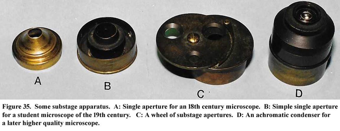
In addition to the mirror, several accessories were available
for the substage. The earliest was a simple set of individual
pinhole apertures to adjust the amount of light and
size of the cone of light (Fig. 35A). Soon, a wheel of
apertures became available; this continued to be supplied
even with achromatic instruments of less than
first class. Later, a single-lensed condenser was used.
Finally, with the development of achromatic lenses,
achromatic condensers of various types were developed (Fig. 35D). These initially were supplied with wheels of apertures; iris diaphragms were not supplied until the end of the nineteenth century.
Polarizers also fit into the substage, which were usually
provided with a method of rotating. Some had
graduated scales (Fig. 13A).
Dark field microscopy could be done using the standard
condenser with dark field stops or with a parabolic
condenser (Fig. 5C).
Another accessory often fitting into the substage area was a dark well holder and dark wells (Fig. 23D). There were used to view small opaque three-dimensional objects in conjunction
with a Lieberkuhn.
OTHER APPARATUS
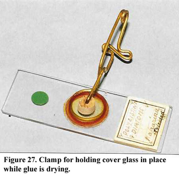
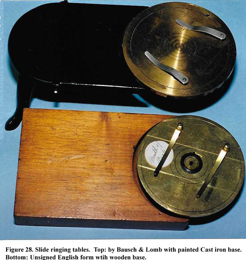
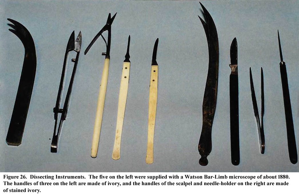
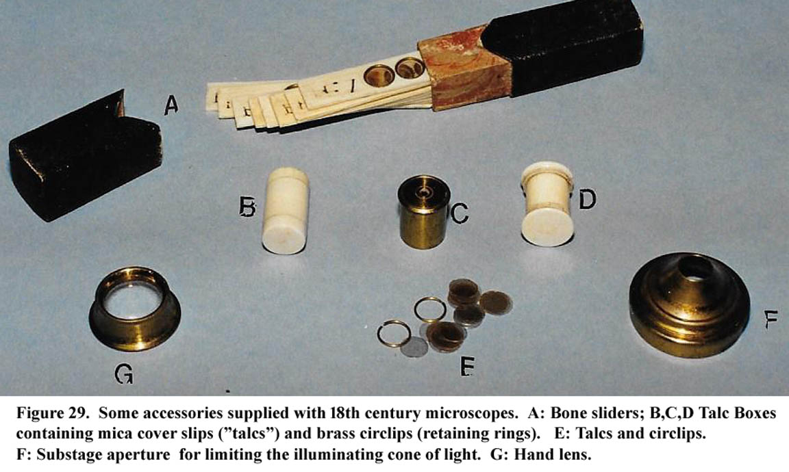
Slide making and dissecting instruments were often supplied (Figs. 26, 27, 28, and 29). Additional accesories related to slide making can be seen on the Collins Slide Kit page.
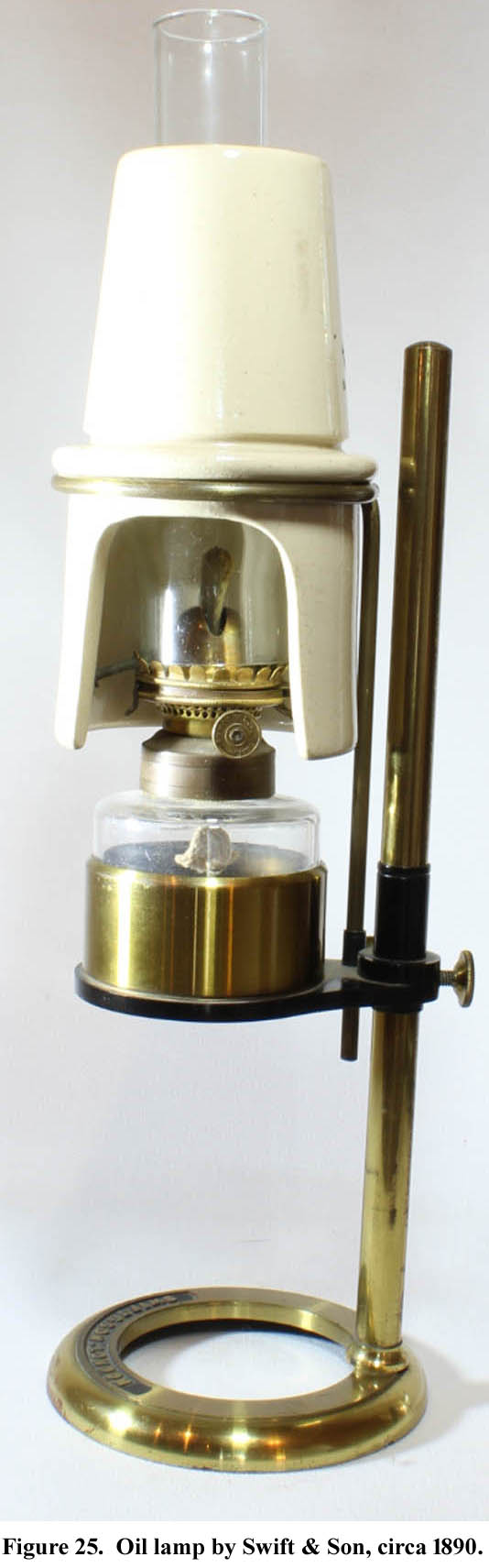
Illumination could be accomplished with light from the sky, a white cloud, a candle, an oil lamp(Fig. 25), or later, gas or carbon arc lamps. To concentrate light, especially for opaque objects, a stand-alone bullseye bench condenser was frequently
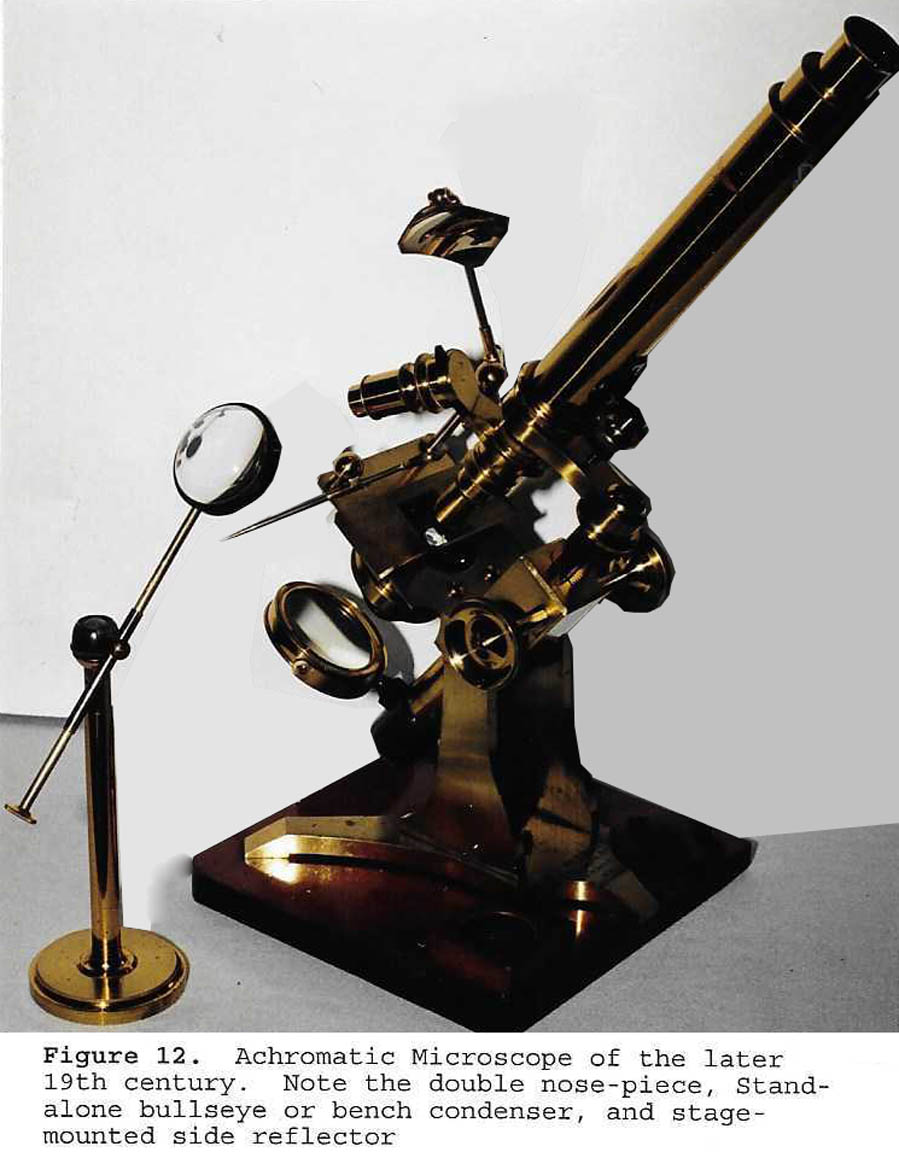
supplied (Fig. 12). Please see the dedicated web article about microscope illumination for more information. Direct sunlight is never used for microscopy as it could cause immediate blindness. The only exception to this rule is when it is used for projection of an image through e.g. a solar microscope.
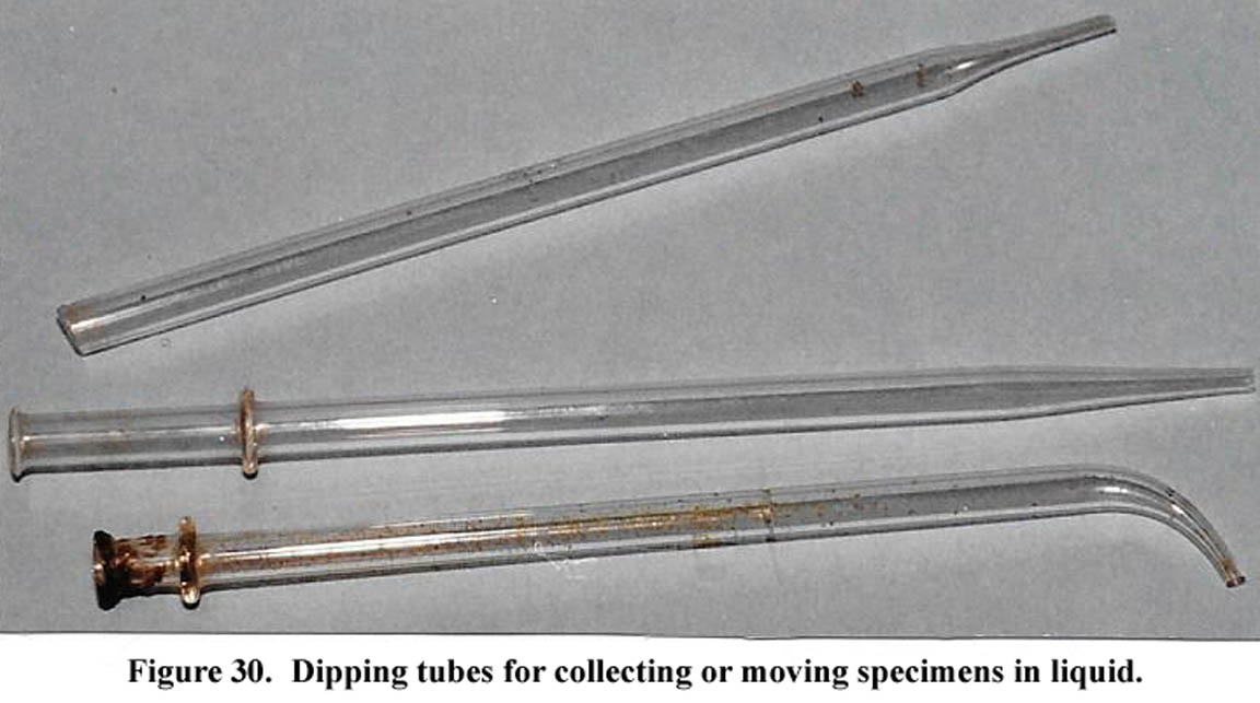 Dipping tubes were used to retrieve microorganisms
from a liquid medium, such as a pond (Fig. 30).
Dipping tubes were used to retrieve microorganisms
from a liquid medium, such as a pond (Fig. 30).
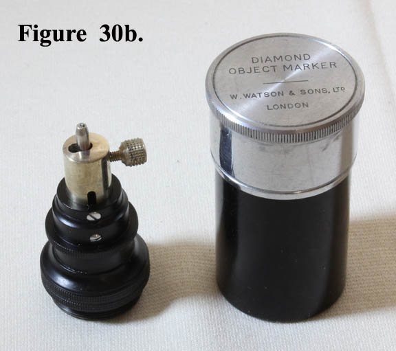 Object markers were used to call attention to a relatively small area of a slide. They commonly came in two forms. Both attached to the nosepiece. One was a simple stamper which could have ink or paint applied and when pushed down onto the slide outlined the area in question. The other type(Fig. 30b), was a tool with a diamond tip, inscribing a circle of adjustable diameter, called a 'diamond object marker'.
FOOTNOTES
1. J. Microscopical Soc of So Calif Vol 3, #4, pp65-79, April 1998.
2I have recently(12/31/16) added the original color images.
3George Adams first produced his 'Universal' Microscope with a rotating wheel of objectives about 1746.
PARTIAL
LIST OF ACCESSORIES SUPPLIED WITH ANTIQUE MICROSCOPES
- Eyepieces
a. Usual types
1)Huygens
2)Ramsden
3)Kellner
4)Orthoscopic (Abbe)
5)Parachromatic (Watson)
6)Holoscopic (Watson)
5)Compensating
b. Micrometer eyepiece
1) Jackson type
2) Ramsden type
3) Fixed micrometer eyepiece
c. Pointer eyepiece
d. Slotted eyepiece to accept slider micrometer, wave plates, etc
e. Variable magnification
f. Strain-free for polarized light
g. Projection eyepiece for photography
h. Projection eyepiece for drawing or group viewing
i. Drawing eyepiece with built-in camera lucida
j. Centering eyepiece with cross-hairs
k. Eyepiece for the determination of grain size in iron-containing metals.
k. Ehrlich Eyepiece for determining the relative ratio of red blood cells to white blood cells on a dry blood smear.
- Eyepiece Attachments or replacements
a. Camera lucida eyepiece cap attachements
b. Polarization analyzer eyepiece cap with calcite prism
c. Tourmaline analyzer eyepiece cap
d. Selenite eyepiece cap adapter
e. Microspectroscope
f. Binocular adapters
g. Pinhole eyepiece
h. Phase telescope eyepiece
i. Double image prism eyecap
j. Wright's Eikonometer for measuring magnifying power of any example of a single eyepiece and single objective.
k. Goniometer eyepiece cap
1)Plain goniometer
2)Leeson Double Image goniometer
l. Dicroiscope apparatus
m. Projection prism eyepiece cap
n. Observation bridge for two observers to view through the same microscope
o. Comparion bridge for viewing images from two microscopes at the same time through one eyepiece, as in ballistics microscopy
- Lister erector lens
- Nosepiece Vertical Illuminators
a. Beck coverslip type
1)Ditto with multiple apertures
2)Ditto with iris aperture and concentrating lens
b. Prism type
c. Stoney large vertical illuminator
d. Electrically illuminated type
- Other Nosepiece attachments
a. Double objective changer
b. Triple objective changer
c. Quadruple objective changer
d. Quick-change and Centering objective changer
e. Rotational attachment (for, or with, analyzer)
f. Analyzers (for polarized light work)
g. Object Markers
1)'Ink Stamp' Marker
2)Diamond Marker
h. Davis shutter nosepiece iris for dark ground work
i. Sliders for slot in nosepiece
1)wave plates
2)quartz wedge
- OBJECTIVES and ATTACHMENTS
a. Achromatic
b. Parachromatic
c. Holoscopic and semi-apochromatic
d. Apochromatic
e. Plan including plan achro and plan apo
f. With correction collar
g. Dual magnification (“Smith’s Quarters”)
h. With built-in prism for vertical illumination of opaque objects
i. Lieberkuhn illuminator objective covers
j. Parabolic illuminator fitting on end of objective
k. Funnel Stops (used inside of objectives for dark ground work)
l. Phase contrast objectives
m. Strain-free objectives (for polarized light work and interference contrast microscopy)
n. Water-immersion objectives
o. Glycerin-immersion objectives
p. Oil-immersion objectives
q. Objective with variable magnification
r. Objective with built-in iris diaphragm for darkground work
s. Smith Interference Contrast objectives with prism on back element
t. Episcopic objectives for dark ground examination of solid objects
- Stage and limb attachments*
a. Side arm illuminators
1) Ross type rectangular parabolic
2) Wenham circular style parabolic reflector with cut-out to allow placement immediately adjacent to the objective
3)Modification of Wenham parabolic reflector incorporating a second small reflector for higher power work.
b. Stage forceps
1) In some cases can be limb
mounted.
2) Often had a sharp spike on one end that could also have a removable black and white disc attached for viewing large opaque specimens.
3) Sometime mounted on a brass or rubber slide
c. Stage 'vice'
d. Stage-mounted or limb-mounted bullseye
condensers
e. Goniometer attachments
1)Adjusting goniometer stage for positioning crystals for study of their interfacial angles with a Leeson Goniometer eyepiece
2)Various forms of Universal stages for measurement of the optical angles of minerals
f. 3-Pronged stage 'mineral' forceps
g. Beck 'Mineral Stage'
h. Fiber analysis stage attachment
i. Black and White disc for stage opening
(older models)
j. Black back-drop slide
k. Needle combs (?)
l. Thin stage replacements for use with
oblique illuminators.
m. Disk Rotator(Beck)
n. Safety Stages
1)Watson Safety Stage
2)Stephenson Type
o. Incubation Stages
p. Hot stages
q. Capillary rotator stage
r. Integrating stages
s. Multiple axis rotation stages for petrographic work
t. Hardness tester
1)The Spencer 'Microcharacter' device
- Special Slides and related
a. Test plates
1)Abbe
2)Diatom test plates with multiple species
3)Test rulings e.g. Grayson or Nobert
b. Test diatoms (single species)
c. Micrometer slides
d. Crosshair slide
e. Maltwood’s finder
f. Selenite slides
1) Stand-alone simple type
2) Inside a brass 'stage' (slide)
3) Mechanical selenite 'stage' (slide) (Leeson Type)
4) Mechanical selenite 'stage' (slide) ) (Hislop Type)
5)Mica-selenite 'stage' (slide)
g. Well slides-single, double, triple, round or oval
h. Black back-drop slide
i. Apertometers
1)Abbe
2)Beck
3)Cheshire Apertometer.
j. Growing Cells
1)Beck Type
- Liveboxes (some with built-in micrometers)
a. Round double chamber on rectangular
brass base
b. Simple Ivory Livebox
c. Brass wet cell slider
d. Round stand-alone double chamber
e. Varley type (wood)
f. Improved brass Varley type
g. Blankley-Rouselet type
h. Merlin improved livebox
i. Beck's Livetrap
- Troughs
a. Glass troughs
1)plain
2)with internal adjustable sprung glass plate and ivory wedge
3)Brass-edged glass trough
- Compressors
a. Lister type first made by James Smith:
spring-loaded with screw adjustment in the
center
b. Lever type -higher pressure
c. Reversible types
1) Piper's
2) Watson's
3) Ross type
d. Wenham’s compressor
e. Rousselet's compressor
f. Parallel plate type (Beck)
g. Barnard's high power compressor (c. 1930)
h. Glass compressors for detection of trichinella
i. Other models
- Animal Restraints (other than compressors)and related items
a. Frog plates
b. Fish plates
c. Fish or eel tubes
d. Coiled wire to extract fish from tube
- Dissection Instruments
a. Scalpels
b. Pins and needles
c. Tweezers and forceps
d. Scissors
- Substage attachments
a. Pinholes adapters with 3 or more aperture inserts
b. Wheel of apertures
c. Achromatic condensers
1)Without additional features
2)With simple wheel of apertures
3)With dark-field apertures
4)With combined aperture wheel
5)With double wheel of apertures
6)With iris diaphragm
7)Kohler type
d. Parabolic darkfield condenser
e. Wenham’s oblique illuminator
f. Substage Amici prism
g. Polarizer
1)Plain type to slip into substage ring
2)Calibrated type which slips into substage ring
3)With swing-out selenites
4)With removable condensing lens on top.
5)'Universal' type with polarizer underneath, condensing lens above, and two wheels for inserts for darkfield work, oblique lighting, mica, and/or selenites
6)Substage Stack of Glass plates polarizer (Powell & Lealand).
h. Dark wells and their holders
Holder can be on a swivel permanently attached under the stage or attachable to the bottom of the stage or into the substage.
i. Selenite substage attachments with polarizer
1)Darker's Stack of 3 rotating Selenites
2)Field's Ratio Micro-polariscope
j. Phase contrast condensers
1)Single phase ring types
2)Heine Type
3)Zernicke Type
k. Interference contrast condensers
l. White Cloud Illuminators
1)Varley type
2)Over-mirror fitting
- Other apparatus on stands
a. Candle stands
b. Screen to protect eyes from stray light
c. Oil or Kerosene lamps
d. Bullseye bench condensers
e. Parabolic rectangular (Ross type) and other side illuminators
f. Gas powered illuminators
g. Carbon arc lamps
h. Electric illuminators
- Dipping tubes
a. Straight
b. Curved
- Camel-hair brush (for brushing fragile specimens onto a slide or for cleaning optics)
- Tools (e.g. for tightening an inclination joint, or the stage retaining ring in microscopes with multiple stages)
- Slide-Preparation Accessories
a.Ringing Tables
b.Warming Tables
c.Coverslip Micrometers
d.Coverslip clamps
e.Smith Mounting Instrument
f.Slide labels and Paper Covers
g.Unused Slides and Coverslips
h.Chemicals including stains, glues and oils
i.Complete slide mounting kits containing most of the above items.
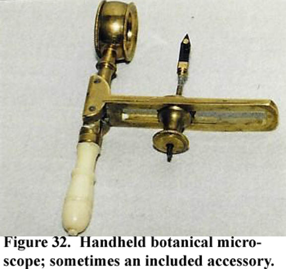
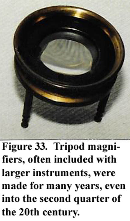
- Hand lenses and simple microscopes
a. Tripod illuminator
b. Multilensed types
c. Folding botanical microscopes
- Bottles For Immersion Oil and Reagent Bottles
a.For immersion oil:
1)plain stoppered glass bottle
2)stoppered glass bottle with glass rod attached to top.
3)stoppered glass bottle with separate glass rod.
4)Bausch & Lomb nickel plated brass (size of objective can).
5)Glass Bottle inside objective case type
a)Bausch & Lomb type
b)Leitz Type
6)Zeiss-Mach type
7)Spencer oil dropper
b. Reagent bottles
1)Glass stoppered bottles (indvidually or in a kit or reagent case
- Illumination Apparatus
a.Candle Illuminator on Stand
b.Oil Lamp Illuminator on Stand
1)With Glass Chimney
2)With Sheet Metal Chimney
3)With Integral Bullseye Lens and/or mirror
c. Monochromatic filters
d. Monochromatic illuminator attachment
e. Electric Illuminators-including carbon arc, mercury lamp, incandescent, and LED lamps
1)On stand
2)Table standing
3)To replace mirror
4)Integrated into base of microscope (modern form)
*Some devices that would traditionally be attached elsewhere, were sometimes made into stages
that would incorporate all parts of the device and would simply be placed on top of the built-in stage. Some examples include a darkfield stage and a selenite stage. The latter was a simple device for elevating the subject slide to a fixed height, providing a shelf below the slide to accomodate a selenite slide, without affecting the height of the subject slide above it.