MICROSCOPE-ANTIQUES.COM © 2013-15.
FROG PLATES AND FISH PLATES OR PANS:
 INTRODUCTION
William Harvey described the circulation of the blood in the year 1615. The one missing link was how the blood got from the arteries to the veins. This was observed under the microscope by Malpighi in 1661 and Antony Van Leeuwenhoek later confirmed these observations in many different sites. For the next century, and beyond, it became common to observe the circulation of the blood through the capillaries under the microscope. This was usually performed in the web of the foot of a frog, or the tail of a fish or eel. For this purpose, frog plates or fish plates (pans) were often standard equipment among the early microscopes. These came in many forms and were made of different materials, including pure glass, metal, and various combinations of brass, glass, wire, and even wood. Adams, used a wire frame. Culpeper and Cuff-type microscopes often had an associated fish pan made of brass. Finally plates made mainly of brass and glass, or rarely, wood were used. Around 1900 these specialized devices became uncommon, as anesthesia made an elaborate restraint system unneccesary. The author is grateful to Dr Jurriaan de Groot for supplying images of three of the plates shown on this page which are in his collection. I am also grateful to the Whipple Museum of the History of Science of the University of Cambridge, for its permission to use its images for educational and nonprofit reference purposes.
FISH AND FROG PLATES
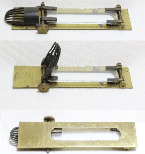
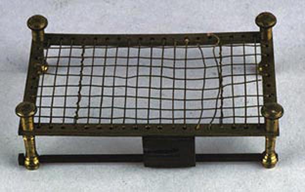 Among the earliest types of apparatus to hold a frog or fish was a metal stage with a wire framework. These would replace the standard stage of the microscope. The images shown to the left show the version sold by George Adams the Elder, as illustrated in his book from the 1750's entitled 'Micrographia Illustrata or the Microscope Explained...' The image to the right of an actual example is from the web site of the Whipple Museum of the History of Science at the University of Cambridge, used for non-profit, educational and reference use only.
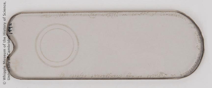 This is a glass form of frogplate, circa 1730, often supplied and signed by Edmund Culpeper with his early versions of Culpeper-type microscopes. This particular example, similar to other known examples, is engraved by Culpeper in his own hand with a diamond pencil with the words:
'This Glasse is to Lay a Fish on to See ye Circulation of ye Blod, ye Animalcula in Liquids or any Transparent objects...'
The image is from the Whipple Museum of the University of Cambridge and used as per their conditions for non-profit, educational use only.
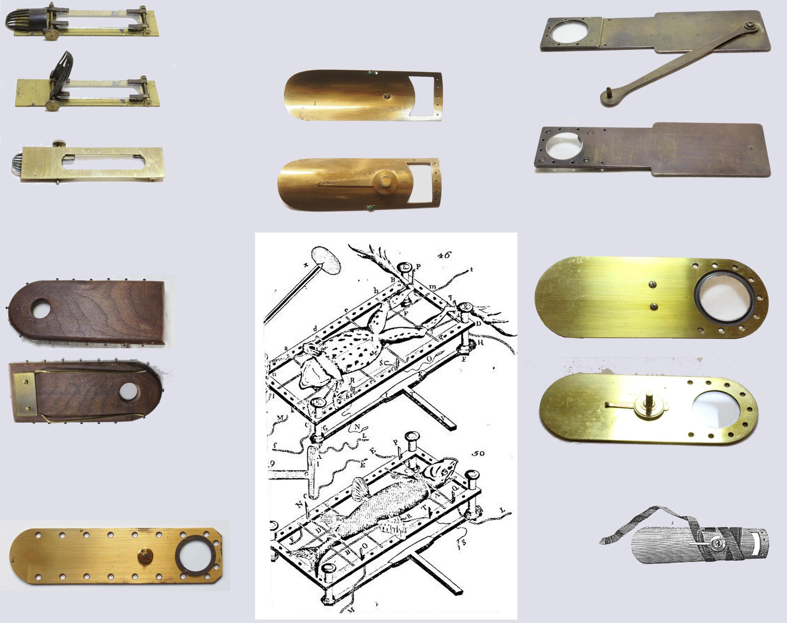
 The next version of this device was the form supplied for many years with Culpeper, Cuff, and Jones type microscopes. These pans are frequently found in the cases of these microscopes in unused condition. They are designed so that when the peg is placed in the appropriate receptacle on the stage, the opening is in the optical axis. These fish plates or fish pans, were a bit smaller than the large frog plates shown below and were mostly used to hold a fish rather than a frog. A ribbon, usually dark green in color, was often supplied to hold the animal in place, and wrapped around the pan. In addition, near the opening for the tail, holes were provided for thread that would secure the tail itself. A peg in the pan allowed it to be mounted in a stage hole. This device was illustrated in George Adams the younger's book, 'Essays on the Microscope' of 1787.
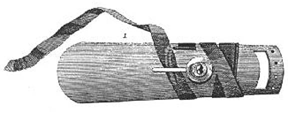
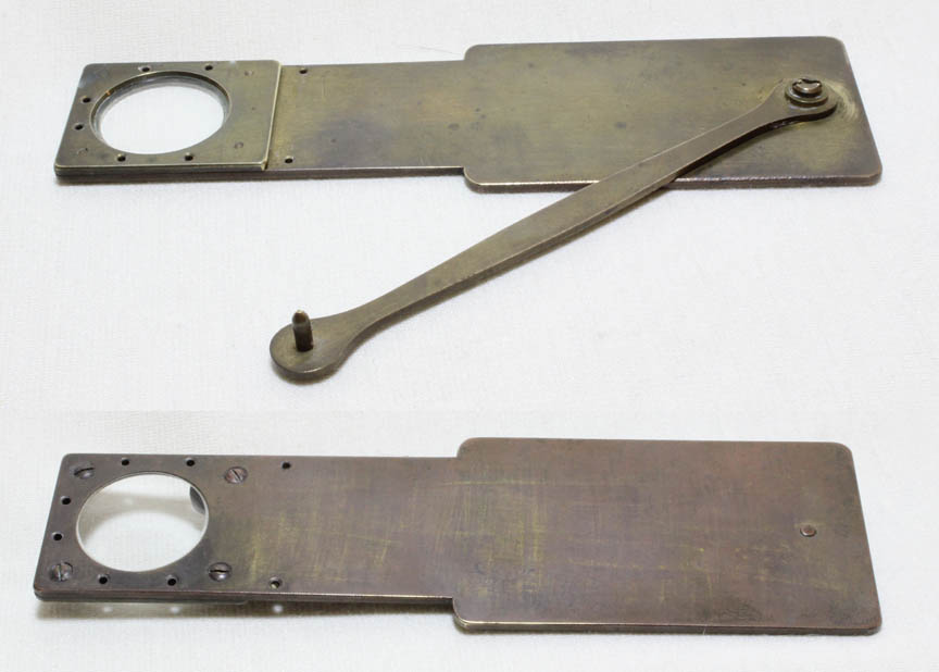
 Starting in the second quarter of the 19th century, thicker and larger supports, supplied for viewing the circulation in the web of the frog's foot were used. The one shown here is from a microscope outfit sold by Varley, but may have been made by Hugh Powell. Also from the collection of Dr Jurriaan de Groot.
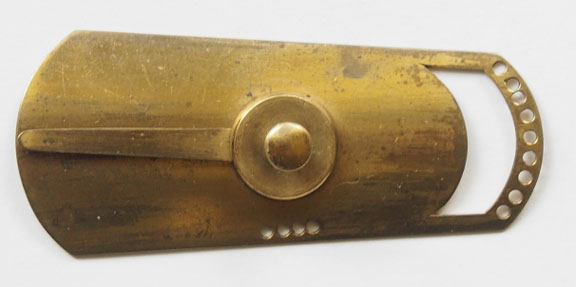
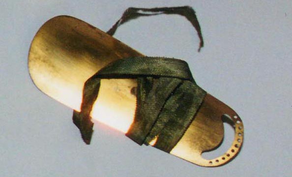
 This example, also from the collection of Dr Jurriaan de Groot, was found with an early Powell & Lealand microscope, and is similar in basic form to the one supplied with the Varley microscope.
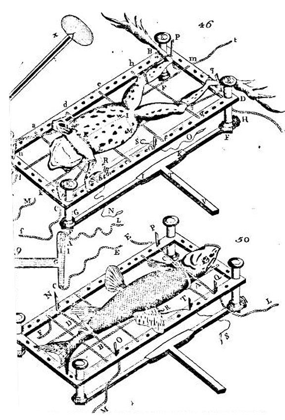
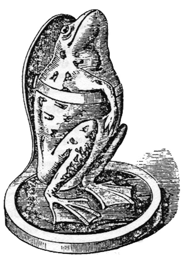 James Parkes and son marketed a frog-holding device which differed from all the others seen on this page. This illustration of the 'Improved Frog Plate,' from 1862, shows the device in use. Using this contraption, made mainly of glass, the frog was anesthetized with ether and then placed vertically with its webs in a more natural position. Two sizes of this device were offered.
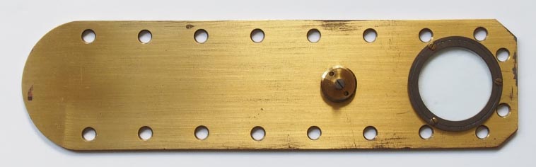
 This is a common type of frog plate from the author's collection, found with Ross microscopes of the second half of the nineteenth century.
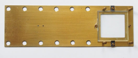
 This rare French form of fish plate was supplied about 1875 with a Alexandre Le Brun a Solar microscope. The narrow but long opening suggests that it might be used to observe more than just the tail of the animal. This plate is the smallest of those shown on this page.
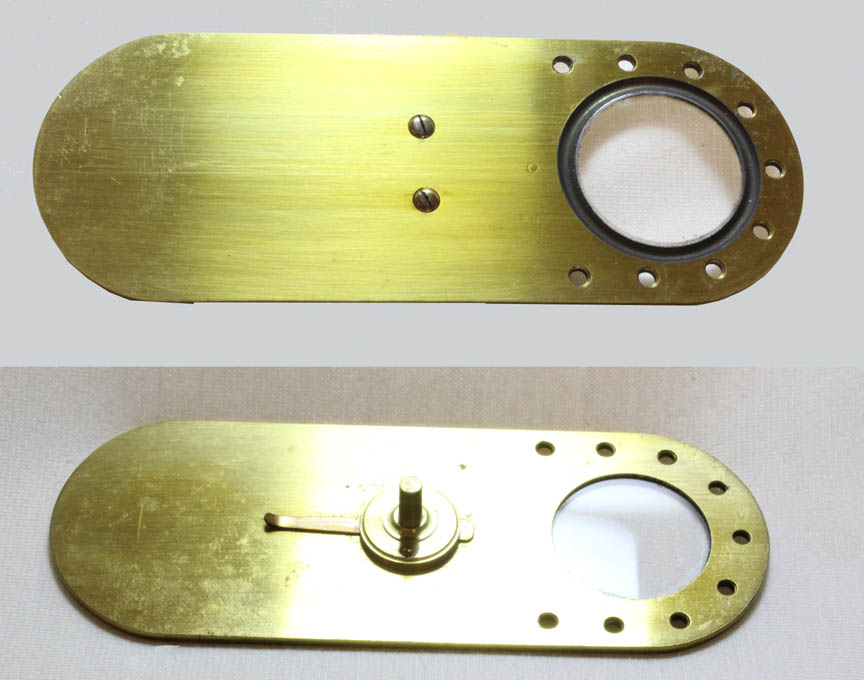
 This is another nineteenth century frog plate, probably of English origin. It allowed more flexible positioning than the others above.
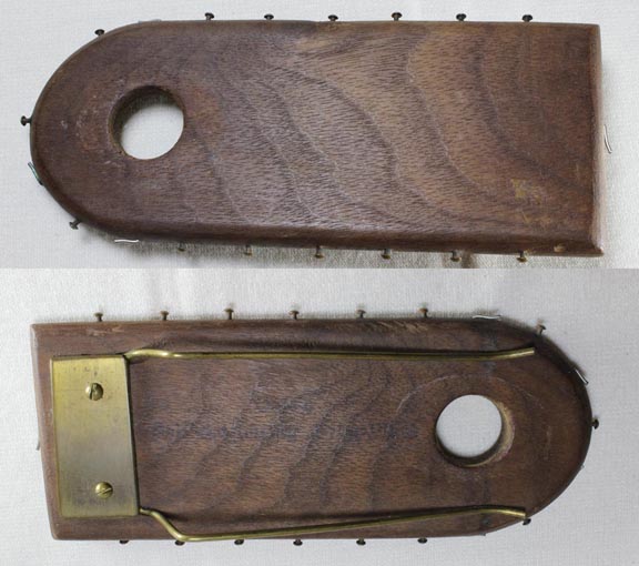 This frog plate, is 'self-adjusting,' and was designed by the American Biology teacher, Aaron Hodgkin Cole. It is mainly made of wood.
The Cole Frog plate page features additional information.
This frog plate, is 'self-adjusting,' and was designed by the American Biology teacher, Aaron Hodgkin Cole. It is mainly made of wood.
The Cole Frog plate page features additional information.