| DESCRIPTION | HISTORY |
Please Click On Any Picture for a Larger Version
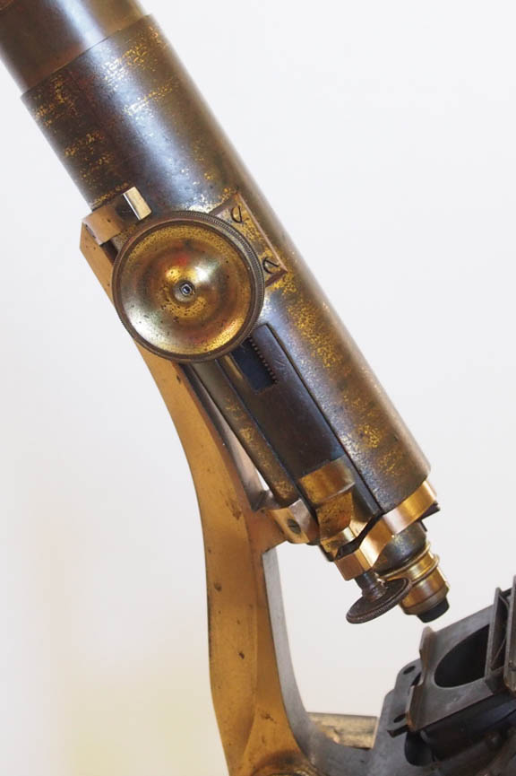
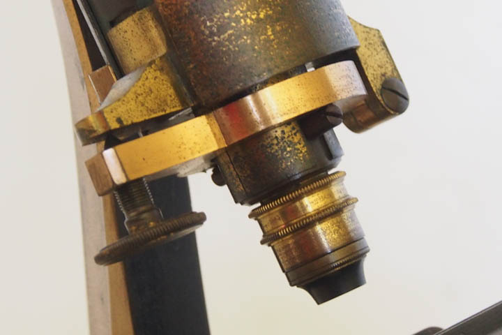
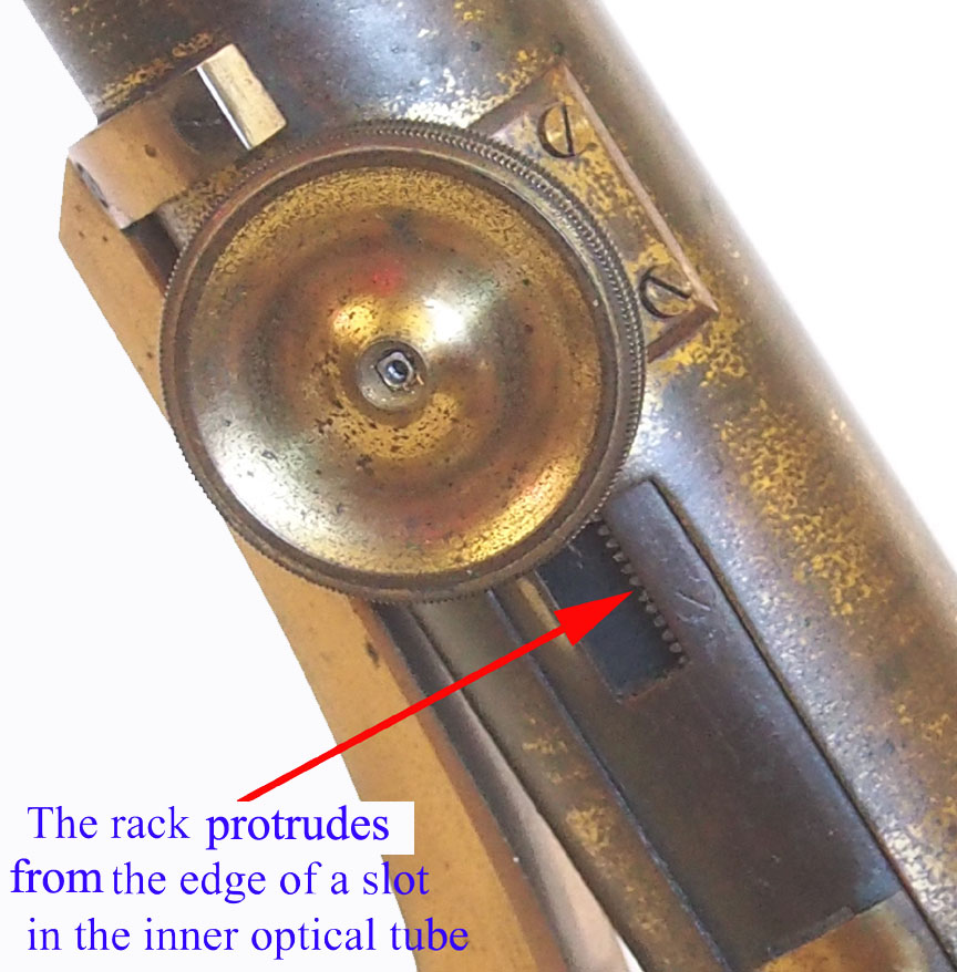 To the top of the limb is attached a fixed external brass body tube with an external dimension of 41 mm. An internal optical body tube slides in this external tube, and has a length of 210 mm and external diameter of 38.6 mm. This inner tube is moved up and down by means of a rack which is screwed into its inner surface with its teeth protruding along a longitudinal slot in this tube, and moved by a pinion attached to a single knurled knob on the right side of the outer stationary tube. According to Paul Ferraglio, who examined one, this arrangement is exactly the same as at least one example of the Powell & Lealand 'Iron Microscope' of almost a decade later.
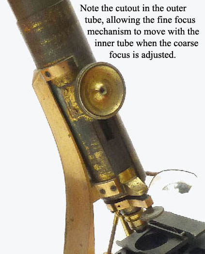
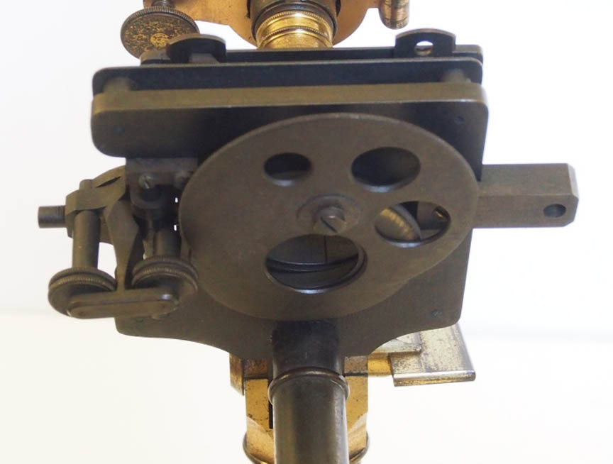
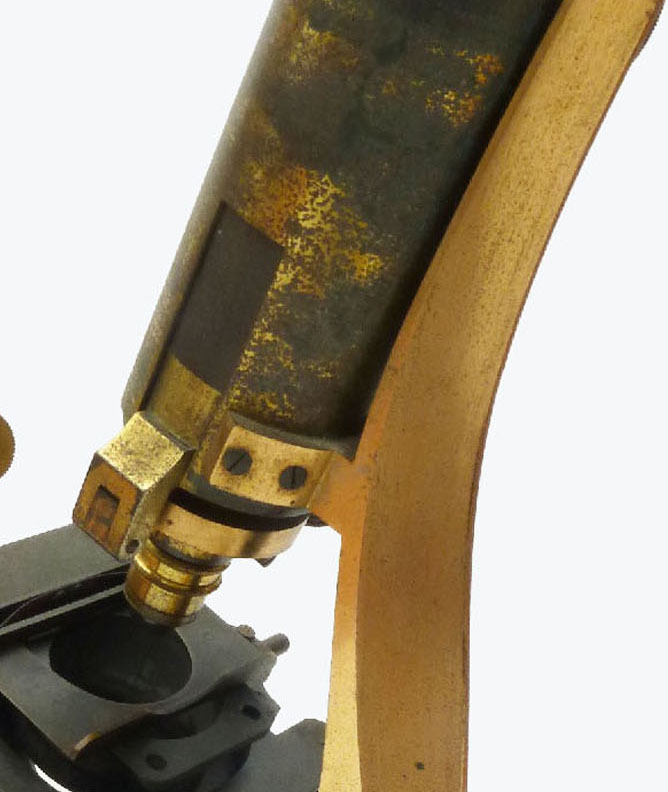 The Ross short lever fine focusing mechanism is positioned at the lower end of the body tube and is adjusted by a knurled knob protruding on the right in the 3 o clock position. A longitudinal recess is cut into both sides of the outer body tube at its lower end to clear the fine focus mechanism during coarse adjustment. This particular coarse-focus arrangement is reminiscent of earlier designs, such as on Martin drum microscopes and precedes the later designs by Ross (as in the so-called 'Penny Cyclopaedia model') where the body tube is fixed to a cradle which moves up and down on a triangular extension of the limb into which the rack is attached. Hugh Powell also utilized the latter arrangement.
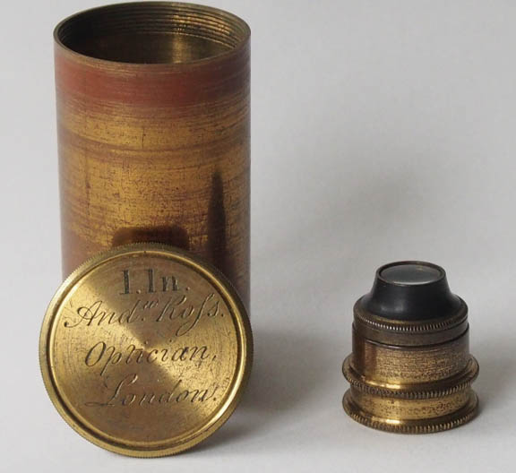 Early Ross 1 inch objective in a can signed 1 In Andw Rofs, Optician, London.
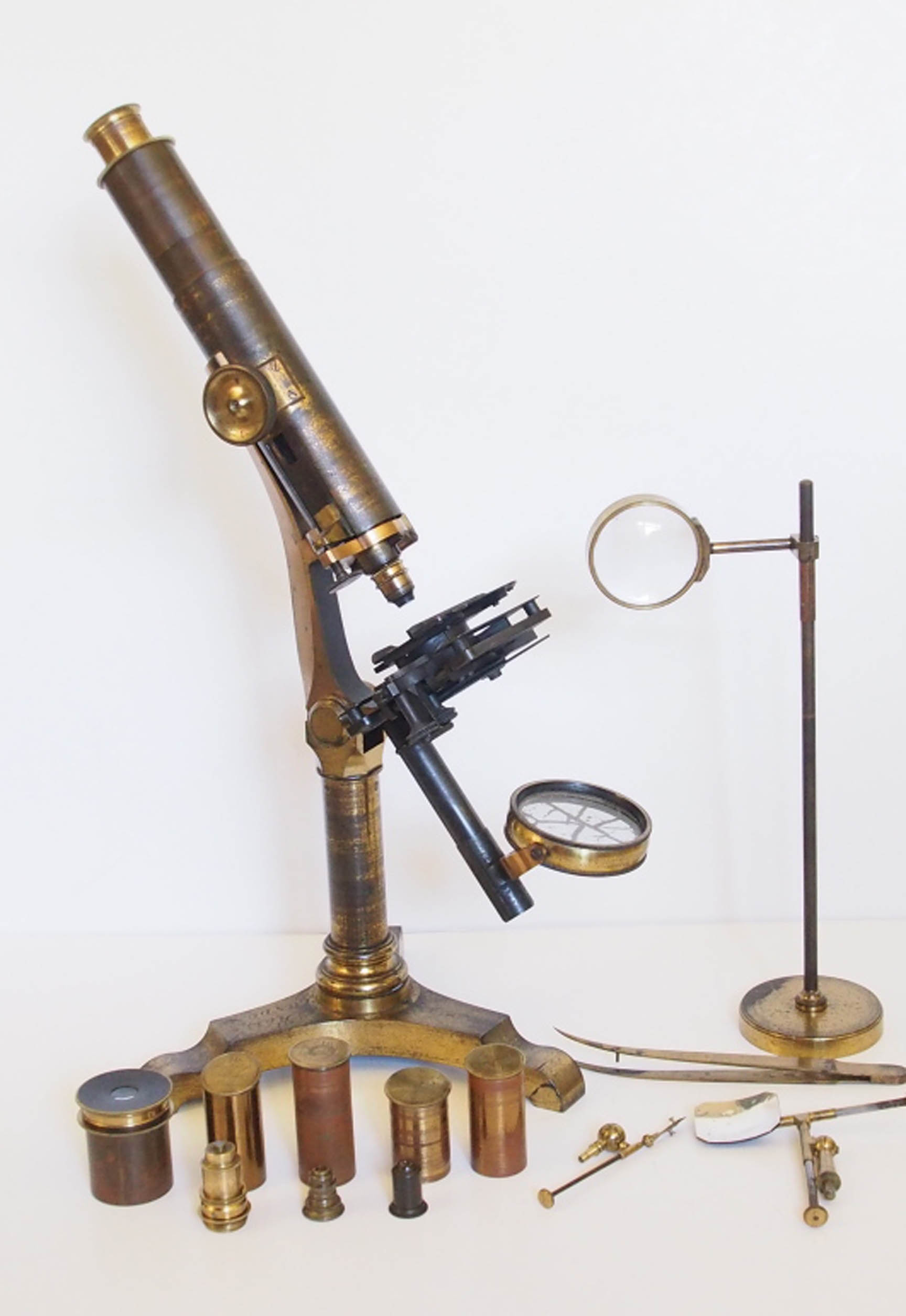
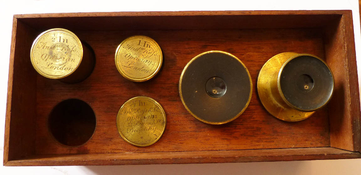
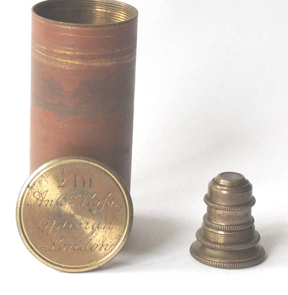
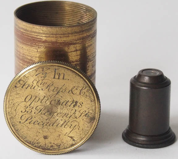 Two early Ross ½ inch objectives, one of these in a can signed ½ In Andw Rofs, Optician, London and one of these in another can signed ½ In Andw Rofs & Co., Opticians, 33 Regent St, Picadilly'
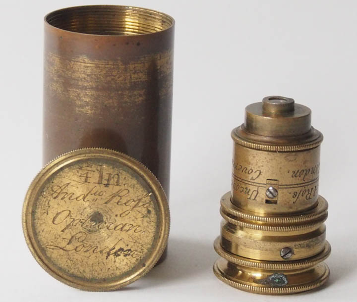 A ¼ inch Ross objective, itself signed 'A. Rofs, London'and also engraved 'Covered' and 'Uncovered' above and below the setting lines, in a can signed: Andw Rofs, Optician, London (later addition) .
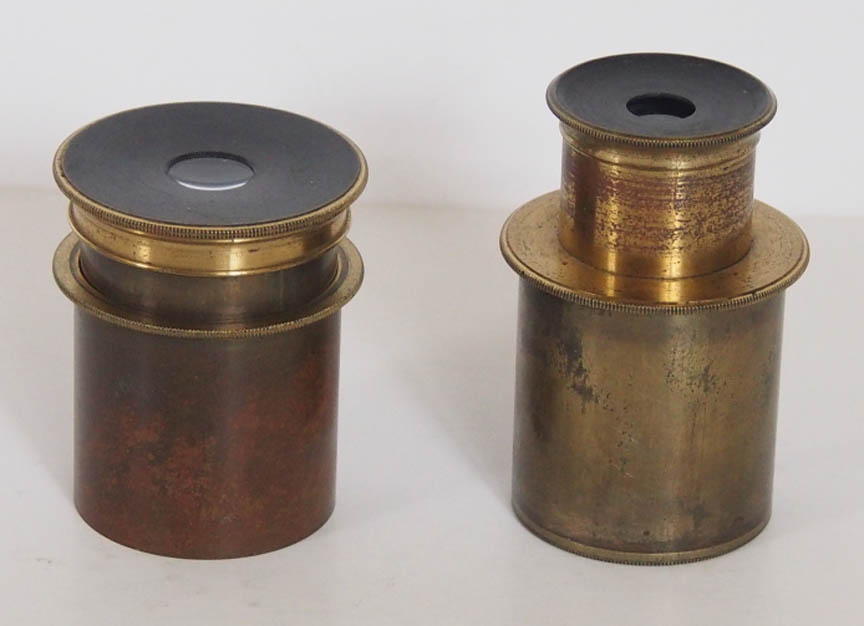 Two eyepieces are included. One is an ordinary 'top-hat' Huygenian type with a power of approximately 8X, and has an outer diameter of 37.4 mm. The second is a Ramsden type, originally a micrometer eyepiece, but lacks its micrometer; this eyepiece allows focusing on the micrometer disc by sliding the top of the eyepiece up and down. As shown here, it was pictured and described by Quekett in the first edition of his Treatise of 1848.
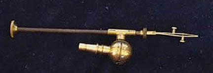
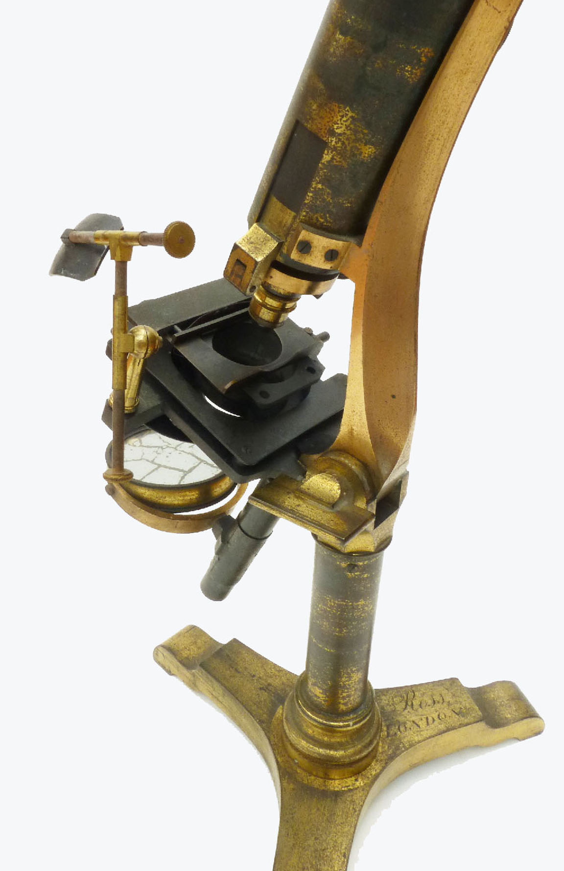
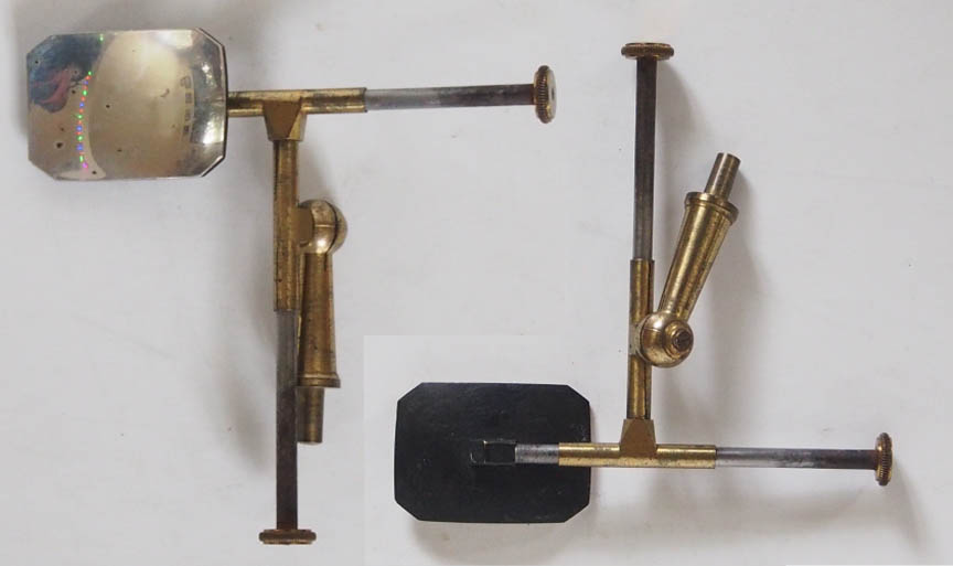 A 'Ross-type' silver side-reflector (probably one of the first ever made see history section) .
 A large pair of brass hand forceps.
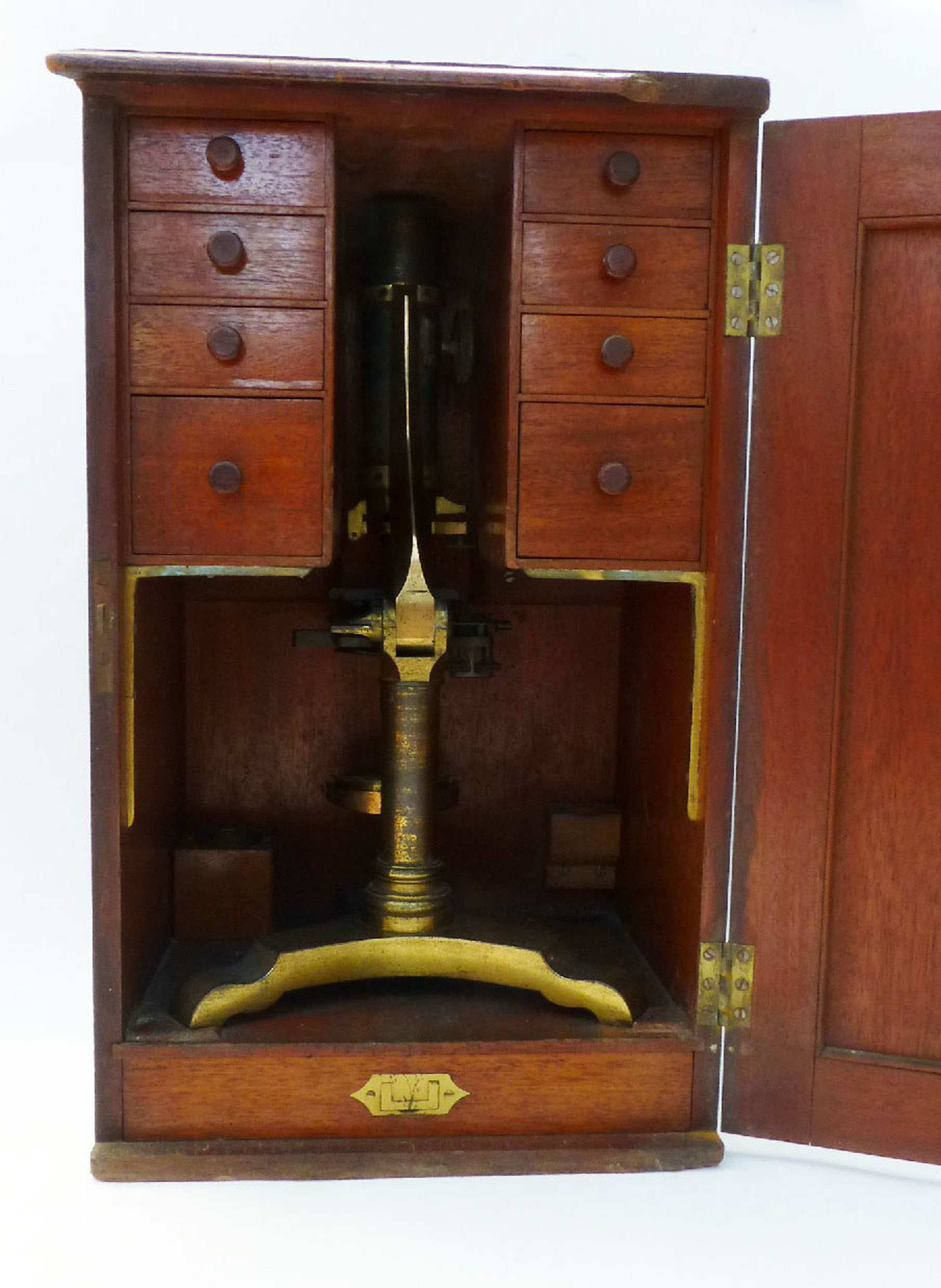
 Free-standing bench condenser on heavy brass foot. The rod can be unscrewed into two halves for storage in the larger center drawer.
 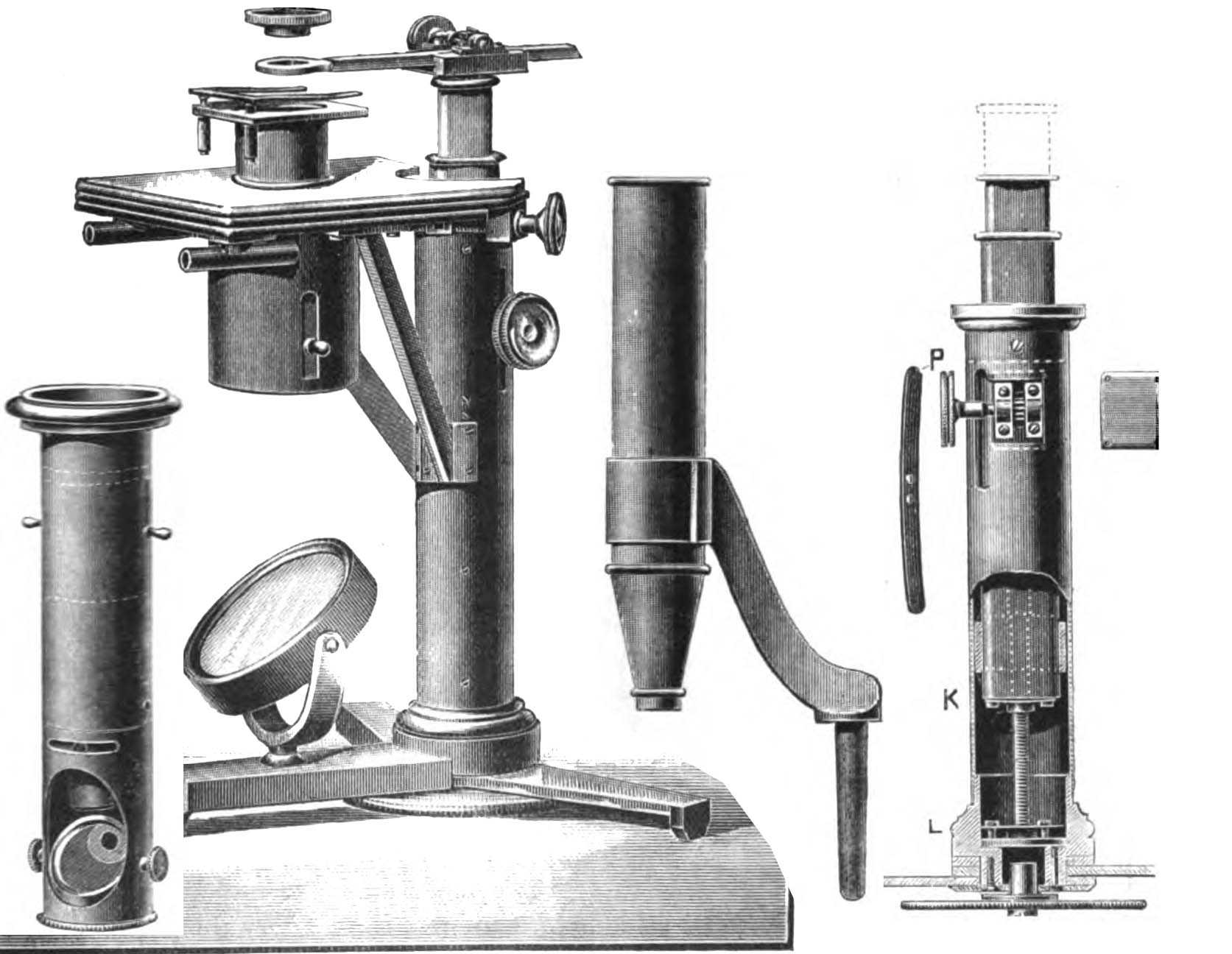 Ross apparently made his first signed microscope stand(left) in 1832 for William Valentine, a plant anatomist of Nottingham. It featured a micrometer fine focus control which was located under the base,or foot, and could be used as either a simple or compound instrument. This period of microscope design, also known as the 'transitional period', during which the first achromatic objectives were fitted to microscopes of an earlier pre-achromatic design, can be rightfully regarded as the crucible from which later more successful designs emanated. Not surprisingly, there was much experimentation by microscope makers who initially worked for the trade, and later started signing their own work. Hugh Powell, Andrew Ross, James Smith, and Andrew Pritchard were part of this environment. The pre-achromatic designs including the Jones most improved, Cary-Gould, Carpenters Improved Compound, and variations on these themes often provided the basis for these experiments aimed at improving both form and function. Many were not signed by their makers, but were signed by retailers such as Bate, Cary, Dollond and Carpenter & Westley. For an example of a transitional microscope of this period, signed by Cary, with features typical of both Andrew Ross and also Hugh Powell, see the Transitional Cary Achromatic Microscope on this site. Note that that instrument has a ball and socket joint at the top of the pillar, similar to many Ross microscopes of the time, and a one-sided gimbal for the mirror, characteristics of the work of Hugh Powell.
 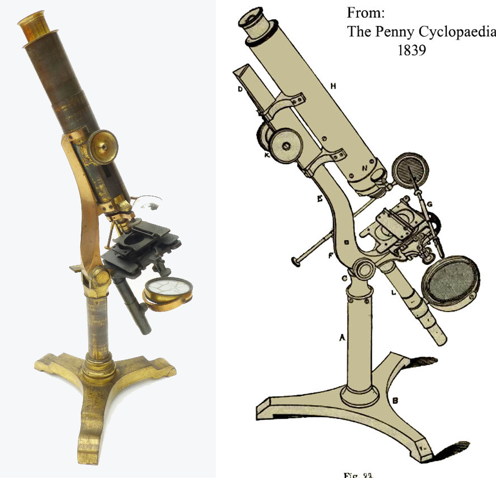 From 1837 Ross traded from 33 Regent Street, Piccadilly as Andrew Ross & Co, Opticians. Until recently, it was always thought that this was in partnership with J.J. Lister, but it has come to light that the partner was in fact one Alfred Aigner, architect and member of the Society of Arts. The dissolution of this partnership was announced in the London Gazette of November 1841 (see Bulletin of the Scientific Instrument Society No 124 March 2015). In Ross microscopes from this period the top of the tripod feet are flat, without the earlier curved ends, which now are simply bevelled downward at about a 45 degree angle. For the first time there is a full Lister limb cast in one piece, with a triangular upper extension into the rear portion of which the rack is recessed.
Hugh Powell also used this type of Lister limb with a triangular profile. (For an example of an early triangular Lister limb microscope attributed to Hugh Powell, see the early microscope Ca 1840 elsewhere on this site). The optical tube is fixed to a cradle, which slides over this triangular bar, and containing the pinion for coarse focus. The short lever Ross nosepiece fine focus is retained on these 'Penny Cyclopaedia models' by Ross. Compare the earlier Ross design featured on this page, with that of the Penny Cyclopaedia model as shown in the images to the left.
From 1837 Ross traded from 33 Regent Street, Piccadilly as Andrew Ross & Co, Opticians. Until recently, it was always thought that this was in partnership with J.J. Lister, but it has come to light that the partner was in fact one Alfred Aigner, architect and member of the Society of Arts. The dissolution of this partnership was announced in the London Gazette of November 1841 (see Bulletin of the Scientific Instrument Society No 124 March 2015). In Ross microscopes from this period the top of the tripod feet are flat, without the earlier curved ends, which now are simply bevelled downward at about a 45 degree angle. For the first time there is a full Lister limb cast in one piece, with a triangular upper extension into the rear portion of which the rack is recessed.
Hugh Powell also used this type of Lister limb with a triangular profile. (For an example of an early triangular Lister limb microscope attributed to Hugh Powell, see the early microscope Ca 1840 elsewhere on this site). The optical tube is fixed to a cradle, which slides over this triangular bar, and containing the pinion for coarse focus. The short lever Ross nosepiece fine focus is retained on these 'Penny Cyclopaedia models' by Ross. Compare the earlier Ross design featured on this page, with that of the Penny Cyclopaedia model as shown in the images to the left. 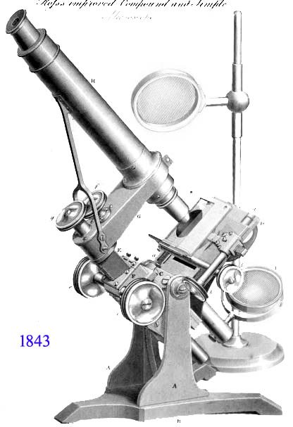 In 1843 Andrew Ross moved to 21 Featherstone buildings, Clerkenwell and the same year saw his announcement of a radical new microscope design as published in the London Physiological Journal of 1843. This would become known as his famous bar-limb model, and set the standard of microscope design for decades to come; it was copied by many other makers. The bar-limb design was also announced only months earlier by Hugh Powell. Powell's design used a different design for the foot however. Andrew Ross died in September 1859, when the firm passed to his son Thomas. It would not be until 1880 before we see a return of a Lister-limb design with the Ross-Zentmayer models.