Microspectroscope
MAKER: Carl Zeiss
Pupillary Micro-spectroscope
c. 1956
DESCRIPTION:
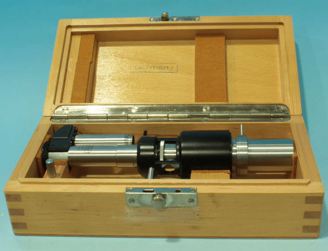
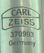 This is a fine working microspectoscope by Carl Zeiss circa 1956. It is signed:
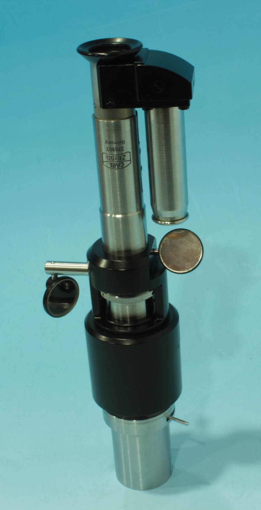
 CARL,ZEISS, 370903, Germany
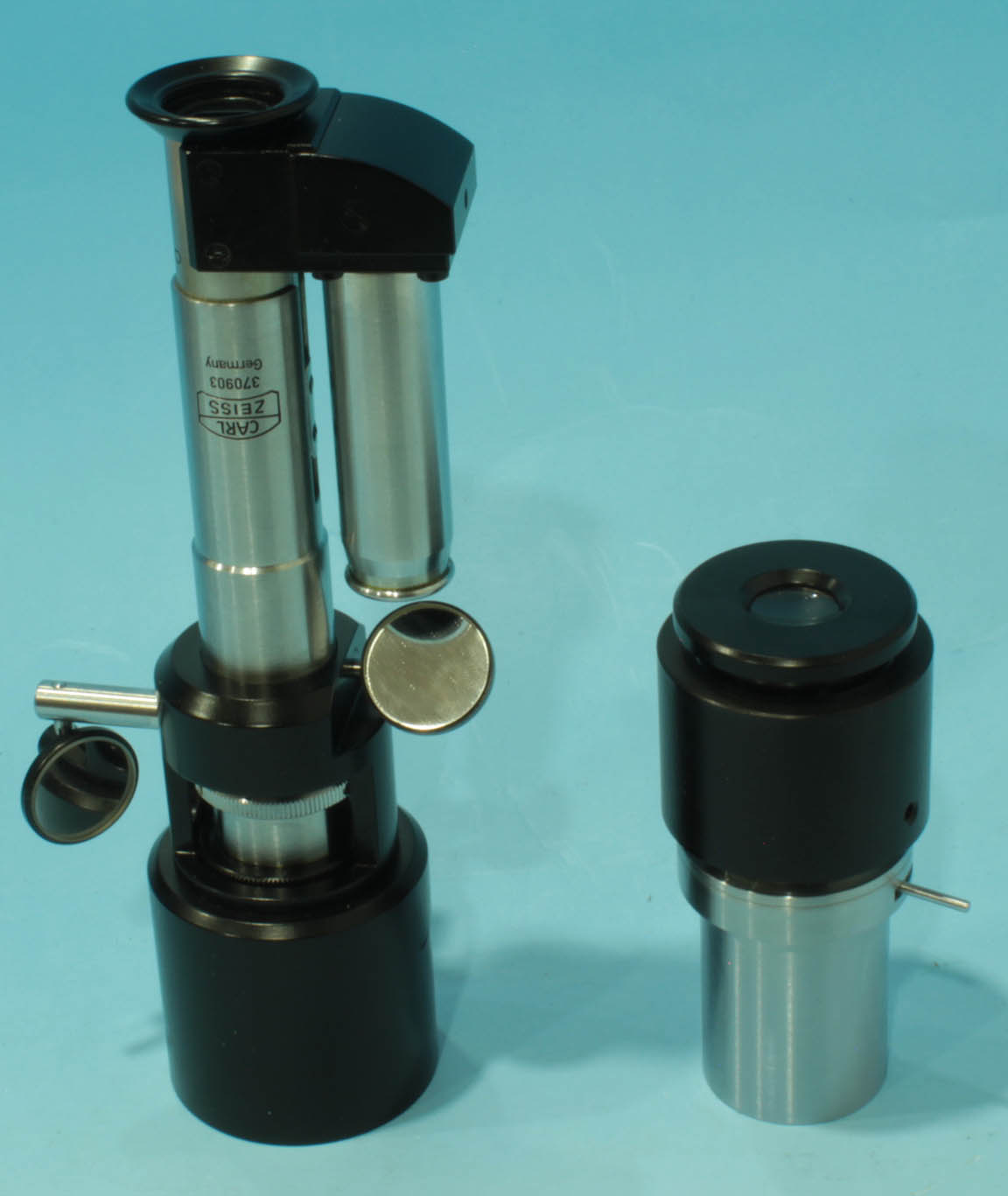
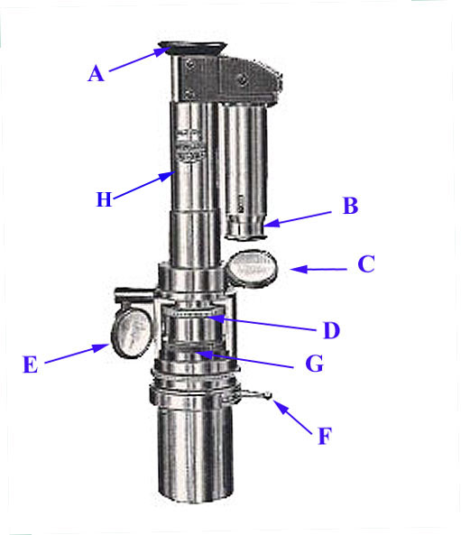
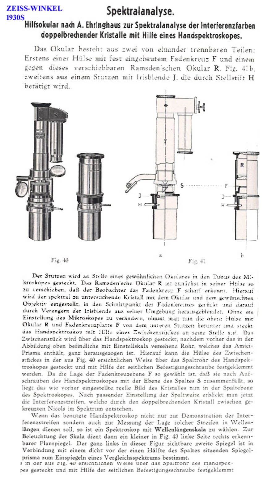
. It is placed in the microscope in place of an eyepiece. It divides into two main parts, the upper part being the spectroscopic optics, and the lower part an eyepiece with an iris diaphragm. It has a mirror (C, below) which is used to direct light to an auxilliary prism projecting a superimposed wavelength scale; This scale can be focused separately using the accessory drawtube(B, below). The mirror E projects light from a reference light source into the device which is seen as a reference spectrum beneath the spectrum of the subject under study. The dial D is used to adjust the slit opening near G. The eyepiece diaphragm is controlled by the lever F. When set up properly, the image of the spectrum under study is shown above the reference spectrum and the wavelength scale is shown superimposed. The scale is not linear, as the wavelengths in the violet end of the spectrum are more spread out than at the red end, and so the divisions get closer and closer to each other as the scale extends from the violet to the red. This results from unequal dispersion of the spectrum due to the action of the prism. Focusing on the subject spectrum is done by pushing or pulling the main drawtube, (H) which is calibrated.
USAGE:
- The subject to be studied was best evaluated in liquid form, therefore it was often disolved in an appropriate solution such as isotonic saline solution, 1% alkali, or glacial acetic acid. This then lent itself to study of very small amounts of a substance. The material might be placed in a livebox for study and therein the thickness of the solution easily varied to optimize the visualization of the spectrum thus produced.
- Moderate intensity lighting of the subject is used
- Another light source of low to moderate intensity is used to illuminate the wavelength scale and provide a comparison spectum, the light for these is reflected in to the device by the two mirrors, C and E respectively.
- The usual eyepiece is used to focus on the specimen preliminarily, and to reduce the diameter of the cone of light of the field seen at the back of the objective by adjusting the aperture diaphragm of the substage condenser.
- The eyepiece of the microspectroscope is pulled out of the bottom of the instrument and is used without the rest of the apparatus to better focus on the specimen, center the specimen, and, by adjusting its diaphragm (F), all extraneous light is cut out; the iris opening can be made very tiny, allowing it to be used for accurate centering.
- After fitting the spectroscope eyepiece back into the sleeve of the spectroscope, and placing back in the microscope tube, the slit width is adjusted by knob D to as thin an opening which still shows shows the spectrum well is found.
- The draw tube on top of the instrument(H) is used to focus as best as possible on the slit and spectrum and the prior step repeated.
- The light from a reference source is reflected into the instrument by adjusting the mirrors which are used to illuminate the scale as well as the reference spectrum oriface.
- The scale focusing drawtube(B) is pushed in or pulled out to bring the scale into sharp focus.
- The result will be seen in the small microspectroscope eyepiece (A).
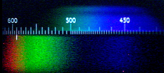
After the above steps the spectrum of the subject being studied will be shown above the reference spectrum, and the wavelength scale will be shown superimposed. In this case the subject is blue-green with wavelengths in the 430-510 Å (angstrom) range.
HISTORY OF THIS INSTRUMENT
The adoption of the spectroscope to the microscope in place of a regular eyepiece was first accomplished by Henry Clifton Sorby using a single prism, but this device, first constructed by John Browning was large and heavy. 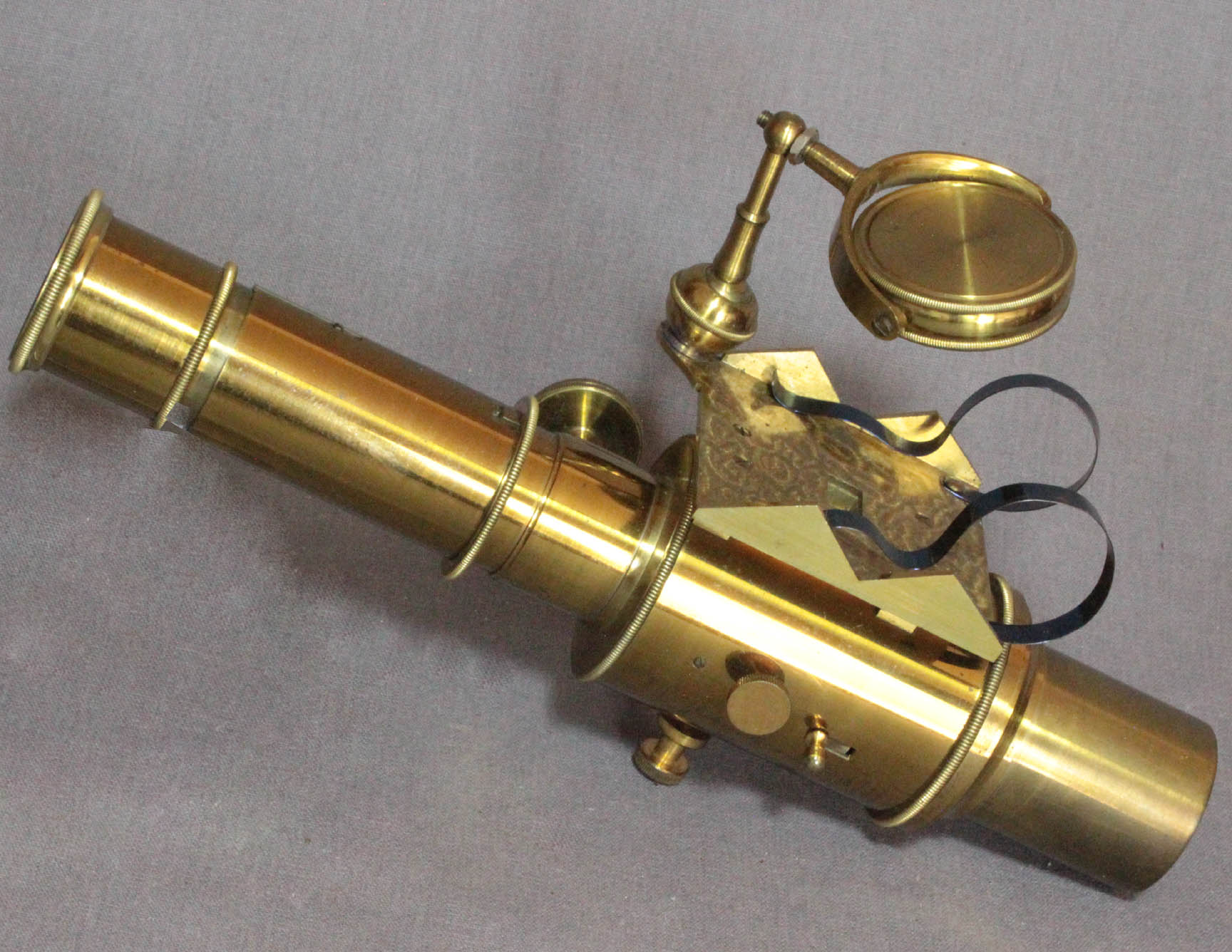 Sorby and Browning then adopted the straight tube prism. This direct vision prism, a cemented chain of three triangular prisms was apparently invented by Amici but was first made by Donati and he published the design in 1862, but again, it was first adapted to the microscope by Sorby and Browning in the 1860s. A better arrangement of 5 prisms invented by Pierre Jannsen in 1877 but again first adapted to the microspectroscope by Sorby and Browning. A 19th Century Browning Microspectoscope is also shown on this site.
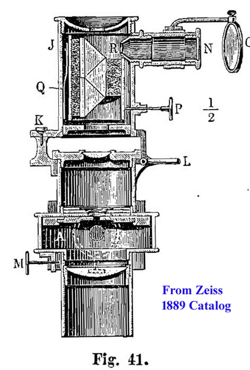
Zeiss sold spectroscopes for use with microscopes starting in the 19th Century. In the 1885 and 1889 catalogs a Micro-spectral Objective after Engelmann
was offered. This device was reported in the Bot. Zeitung of 1882 No 26 and Pfluger's Archive, Bd XXVII p 464 and Bd XXIX p 415. This device projected a spectrum on the the object on the stage from below it, hence was a substage device.

But in the same years, Zeiss offered an Abbe-type
microspectroscope finished in lacquered brass. This Sorby-Browning type of microspectroscope was not an Abbe invention, being made earlier by Browning and designed by Sorby.
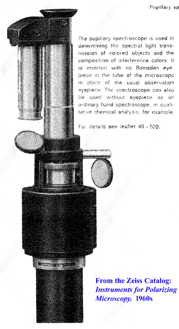
The type of Pupillary
microspectroscope featured at the top of this page was sold by Zeiss from the 1920s through the 1960s.