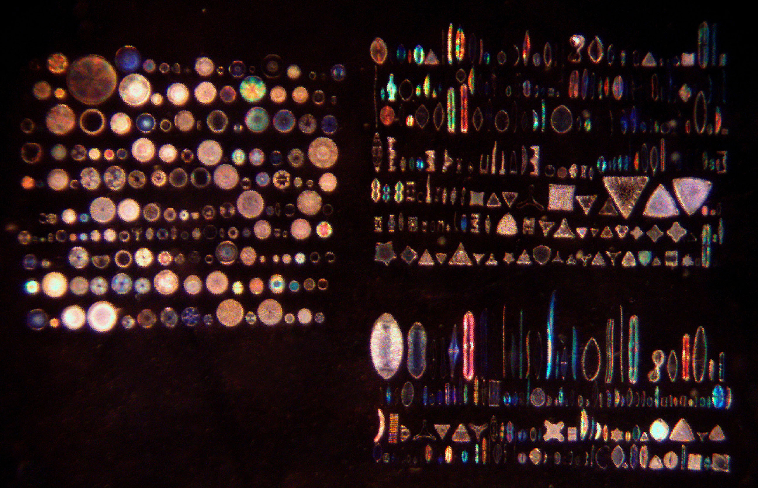
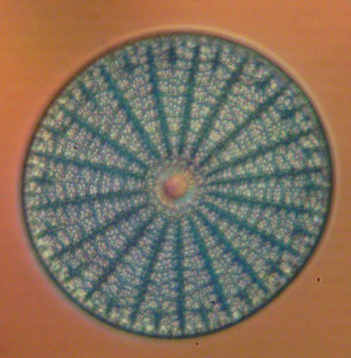
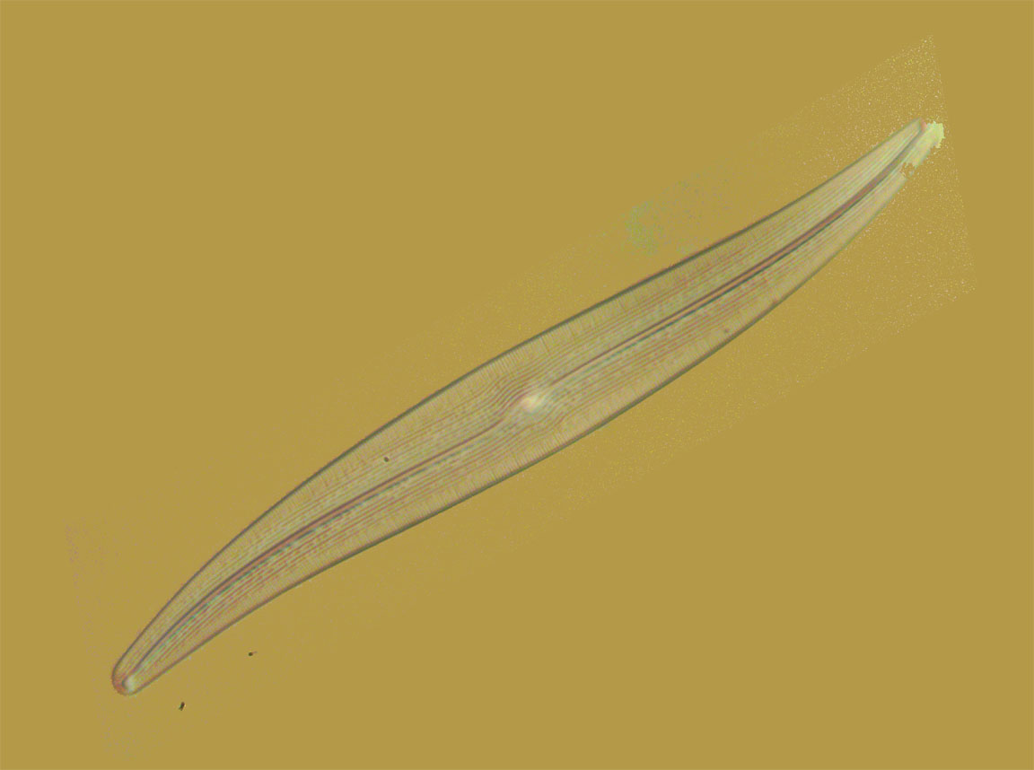
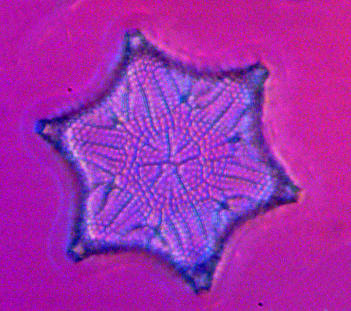
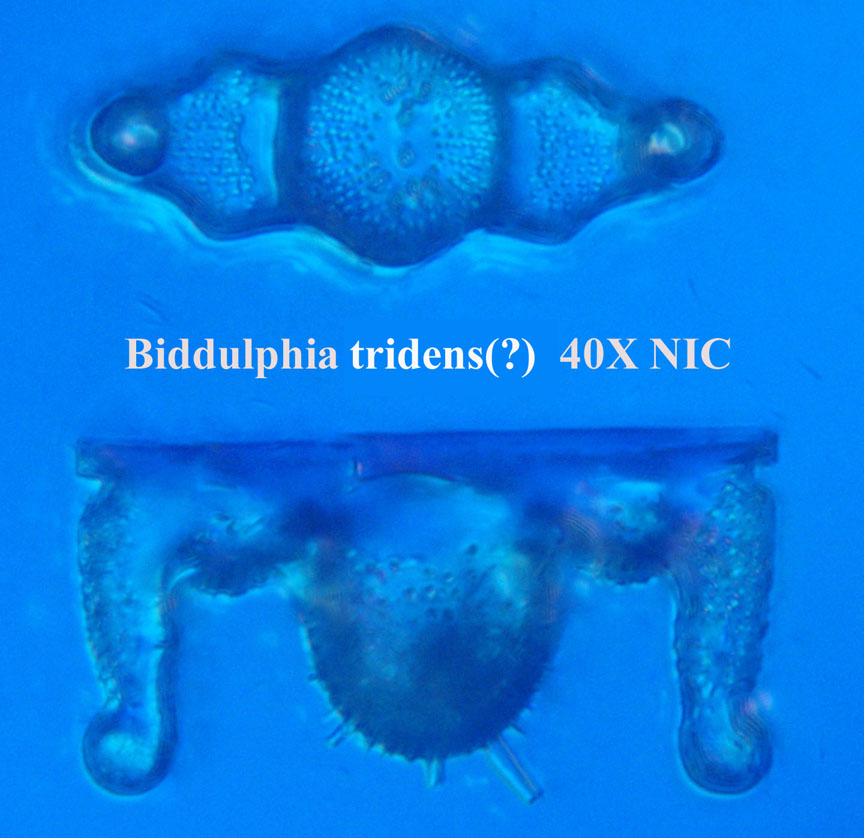
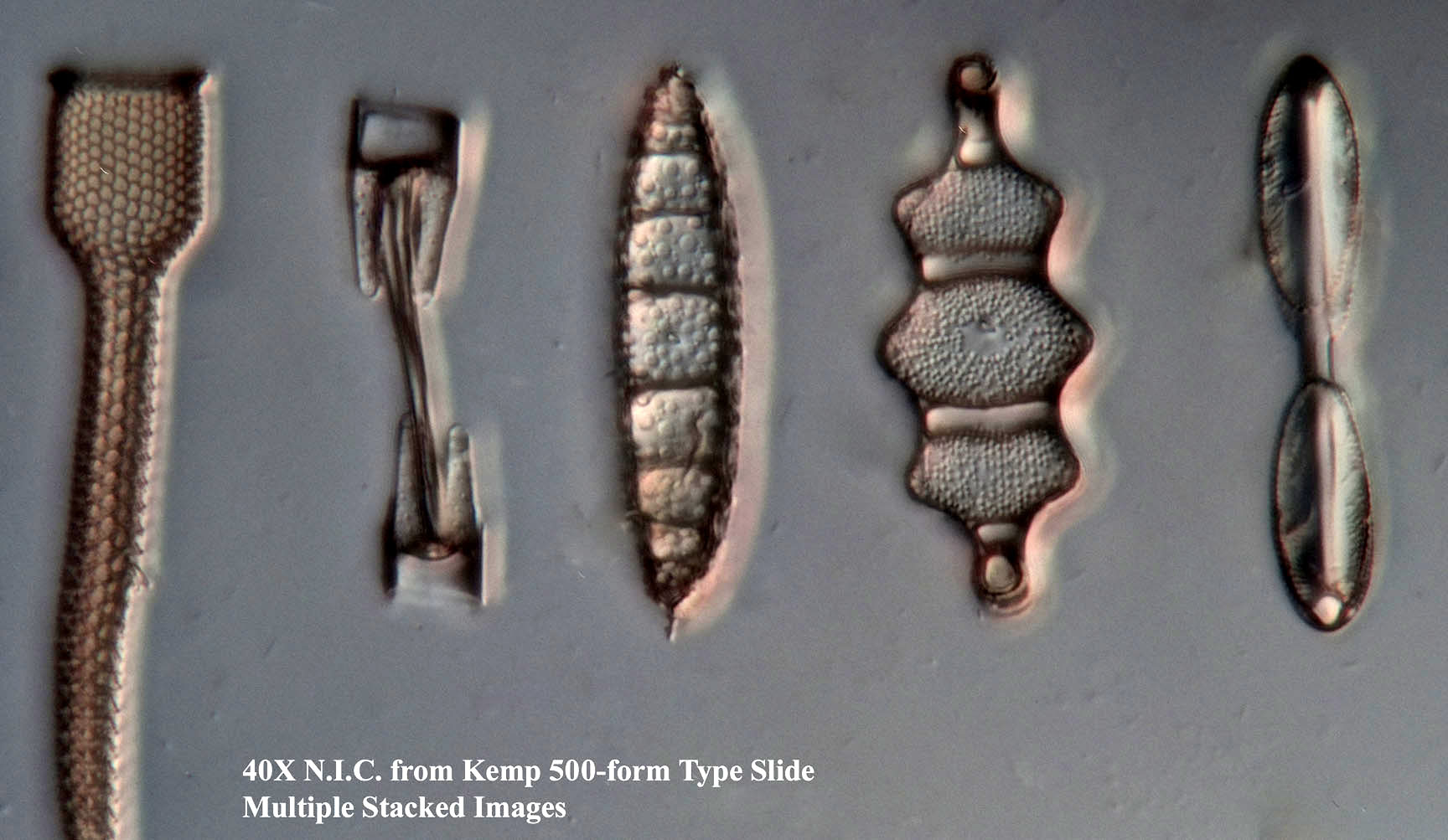
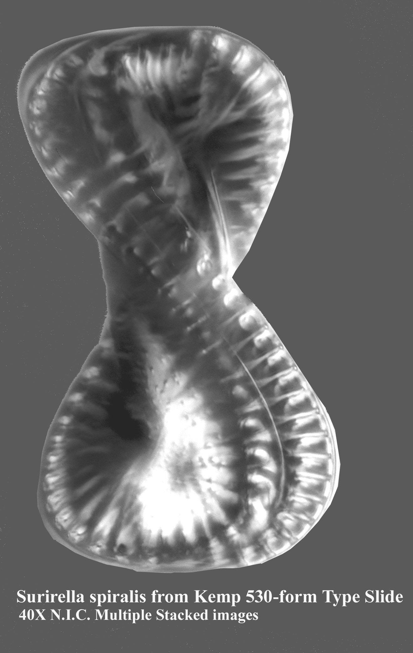
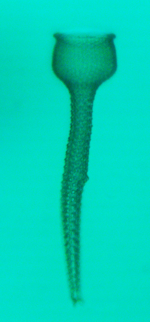
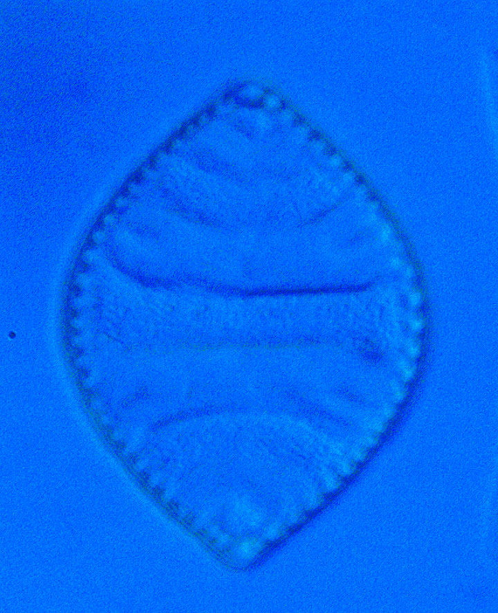
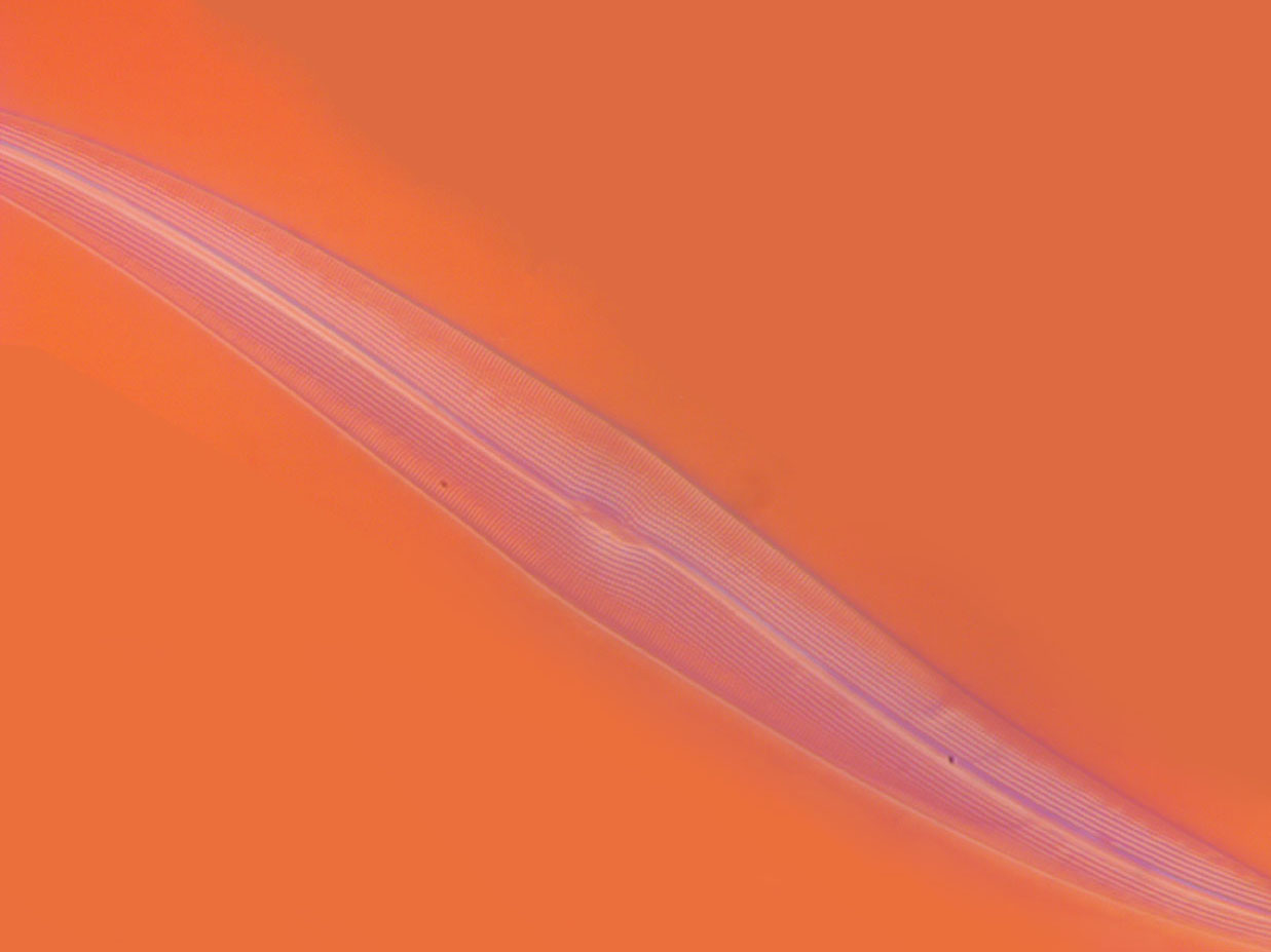
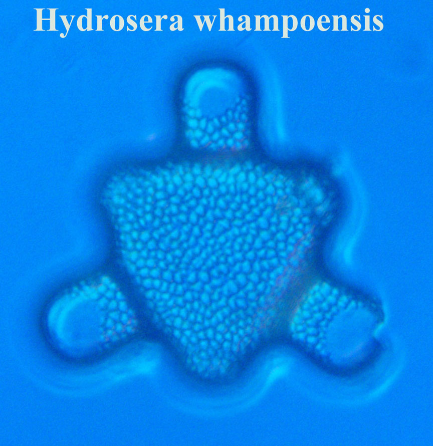
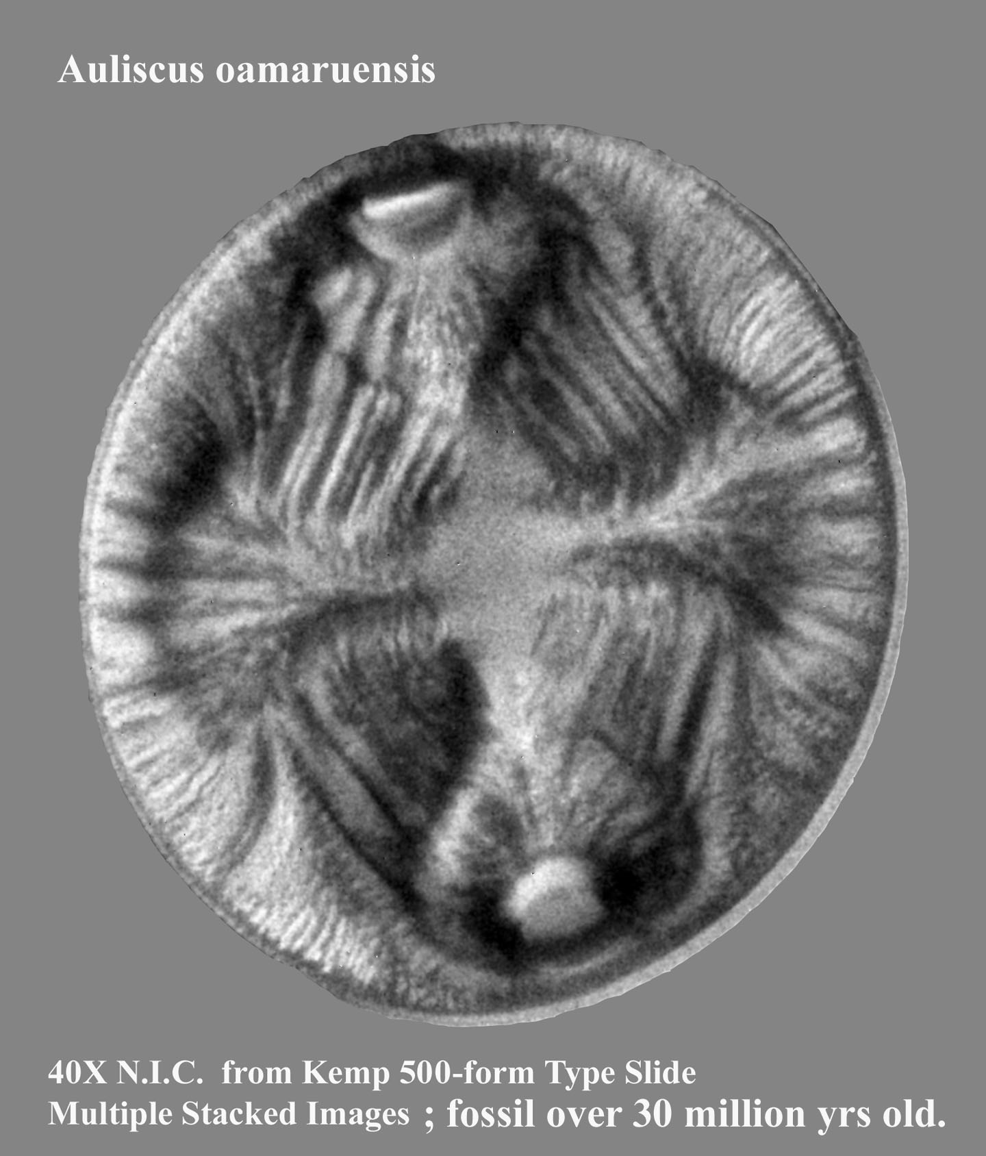
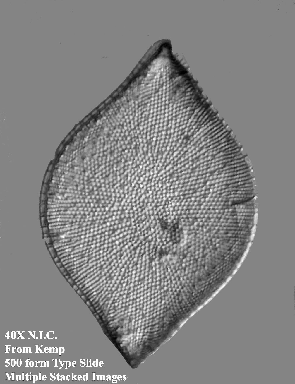
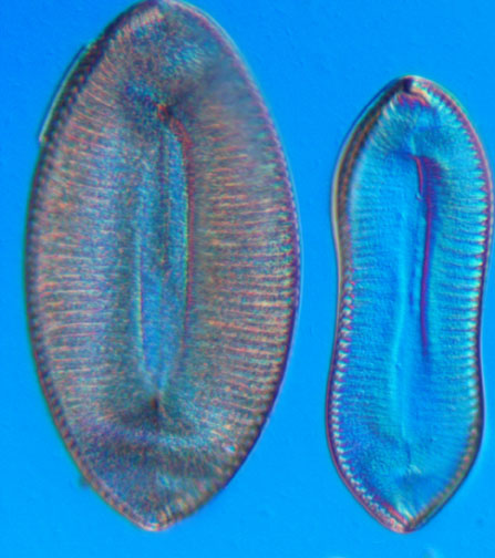
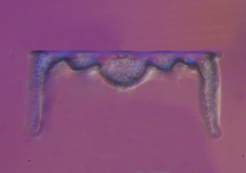
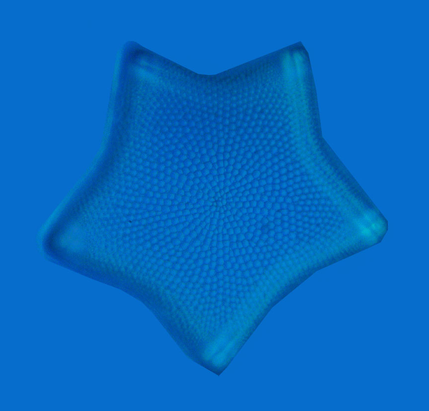
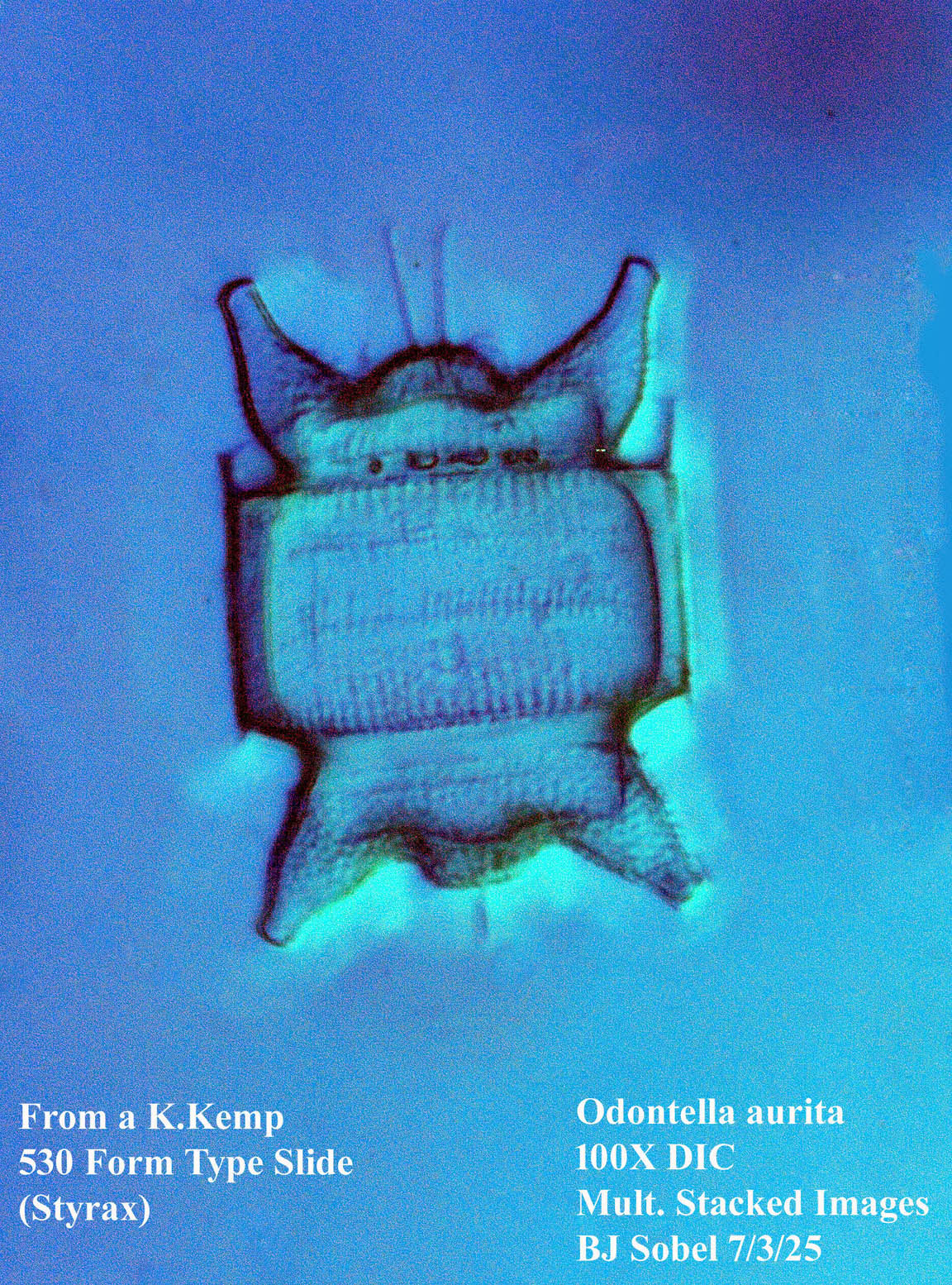
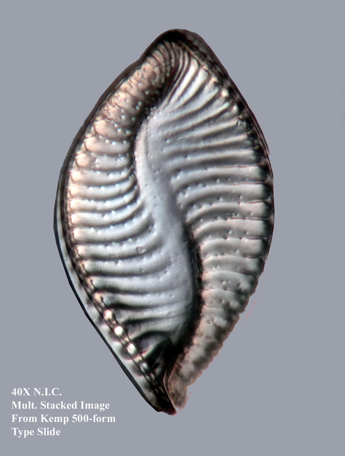
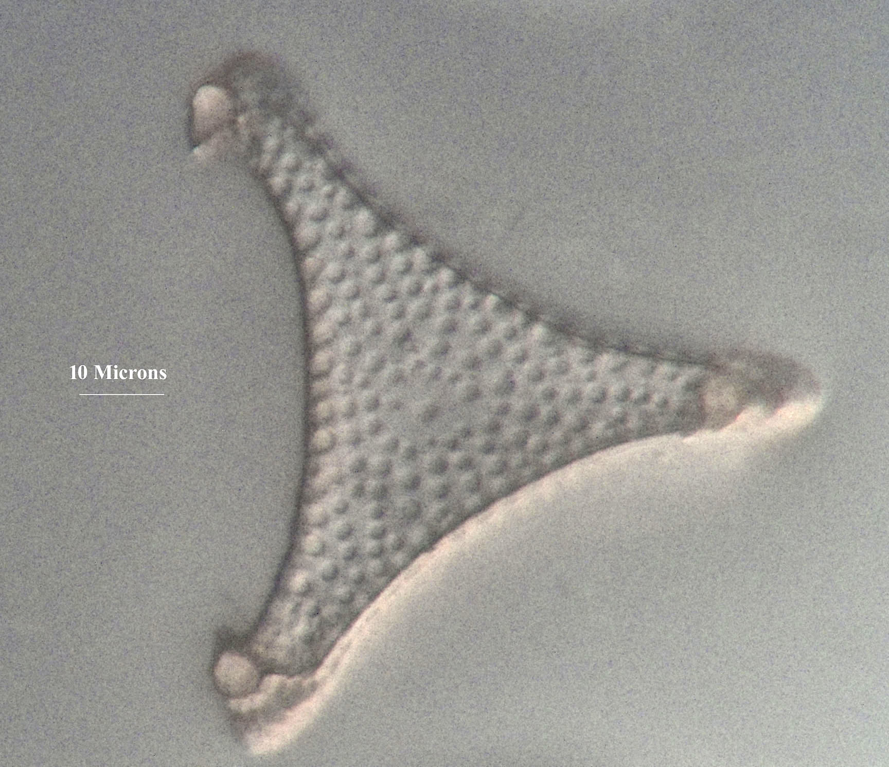
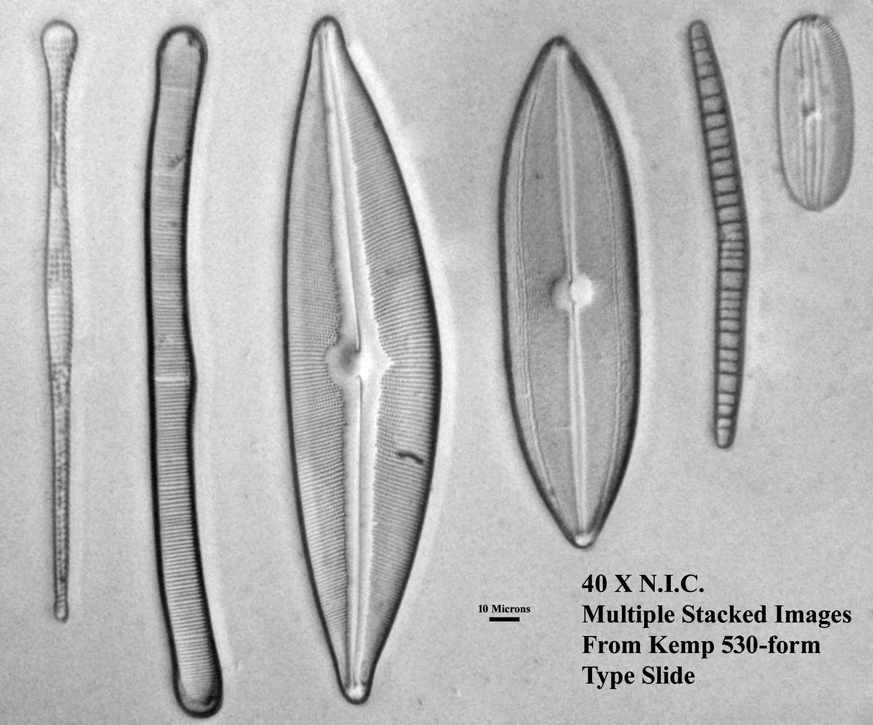
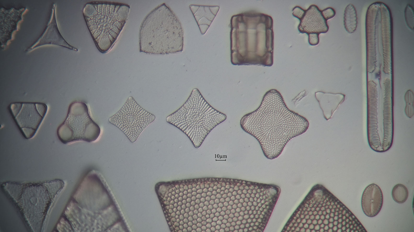
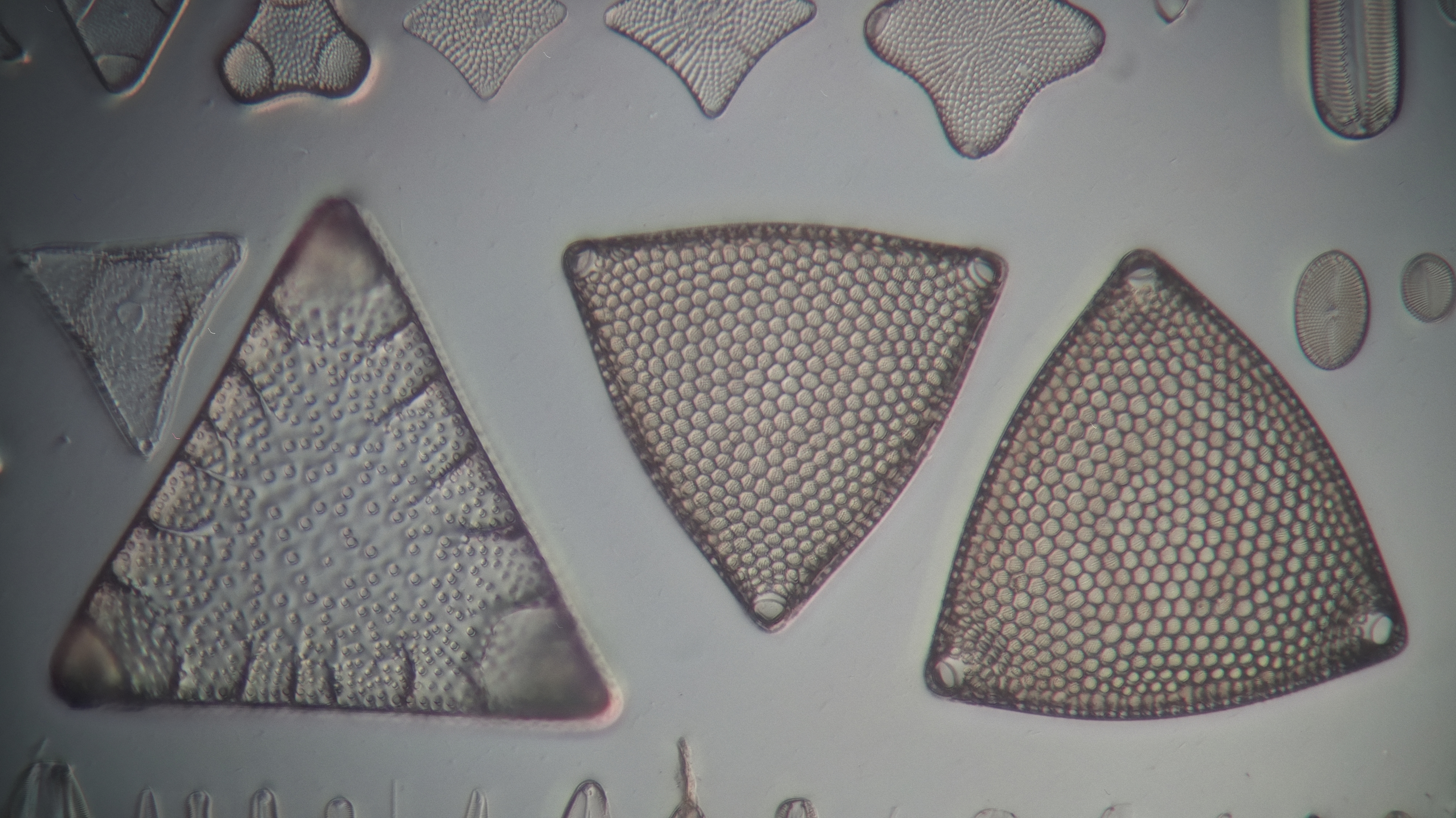
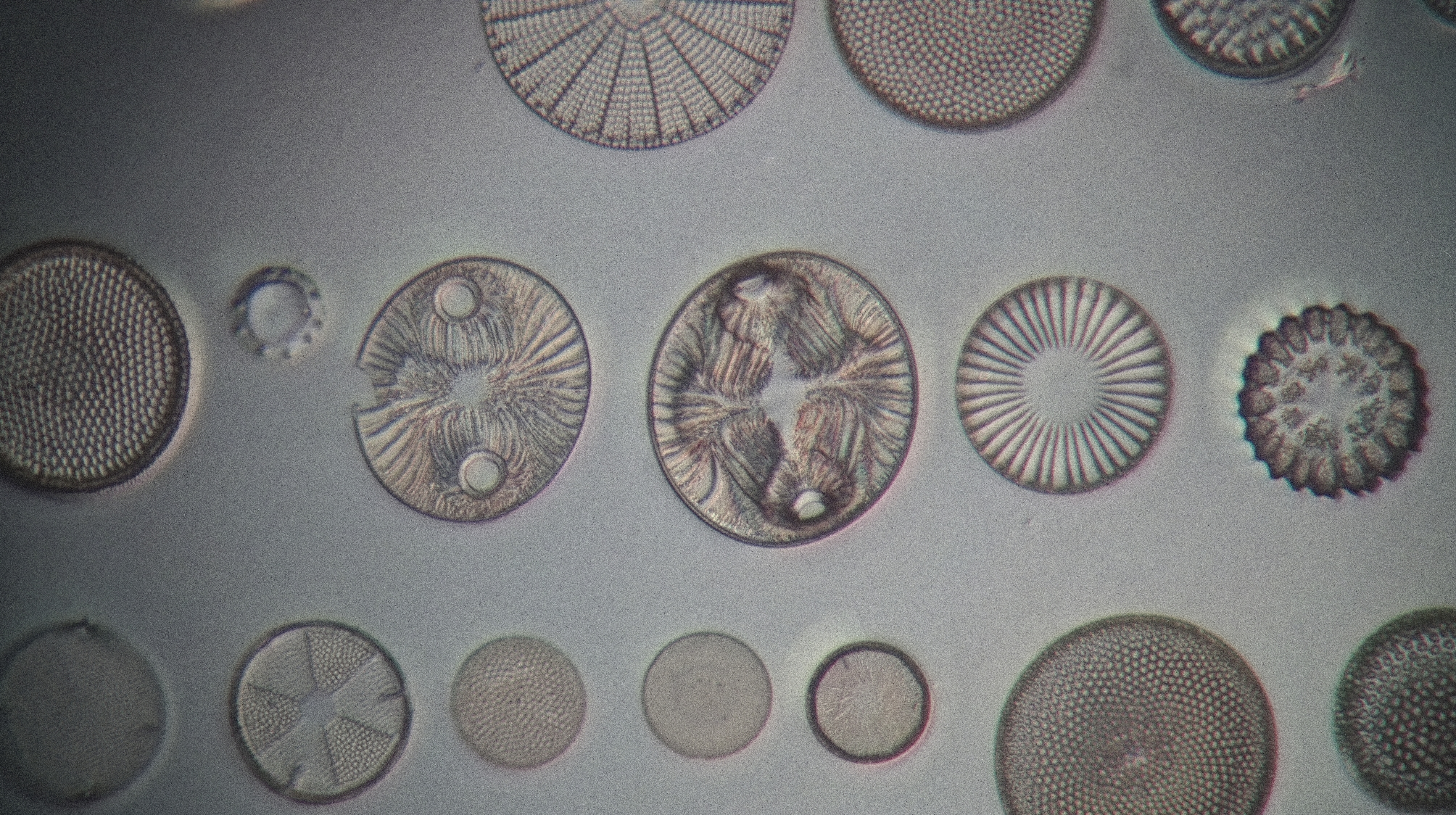
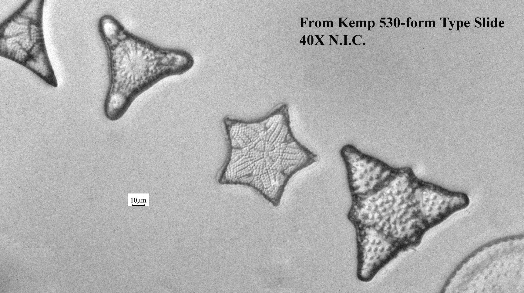
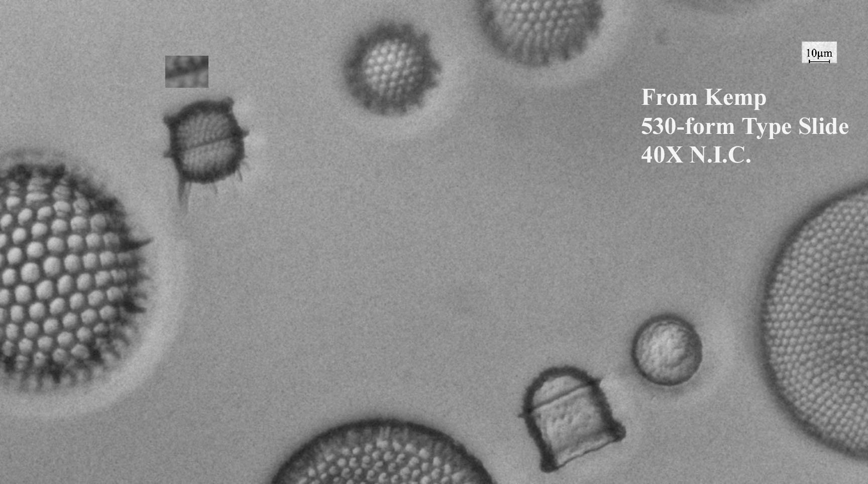
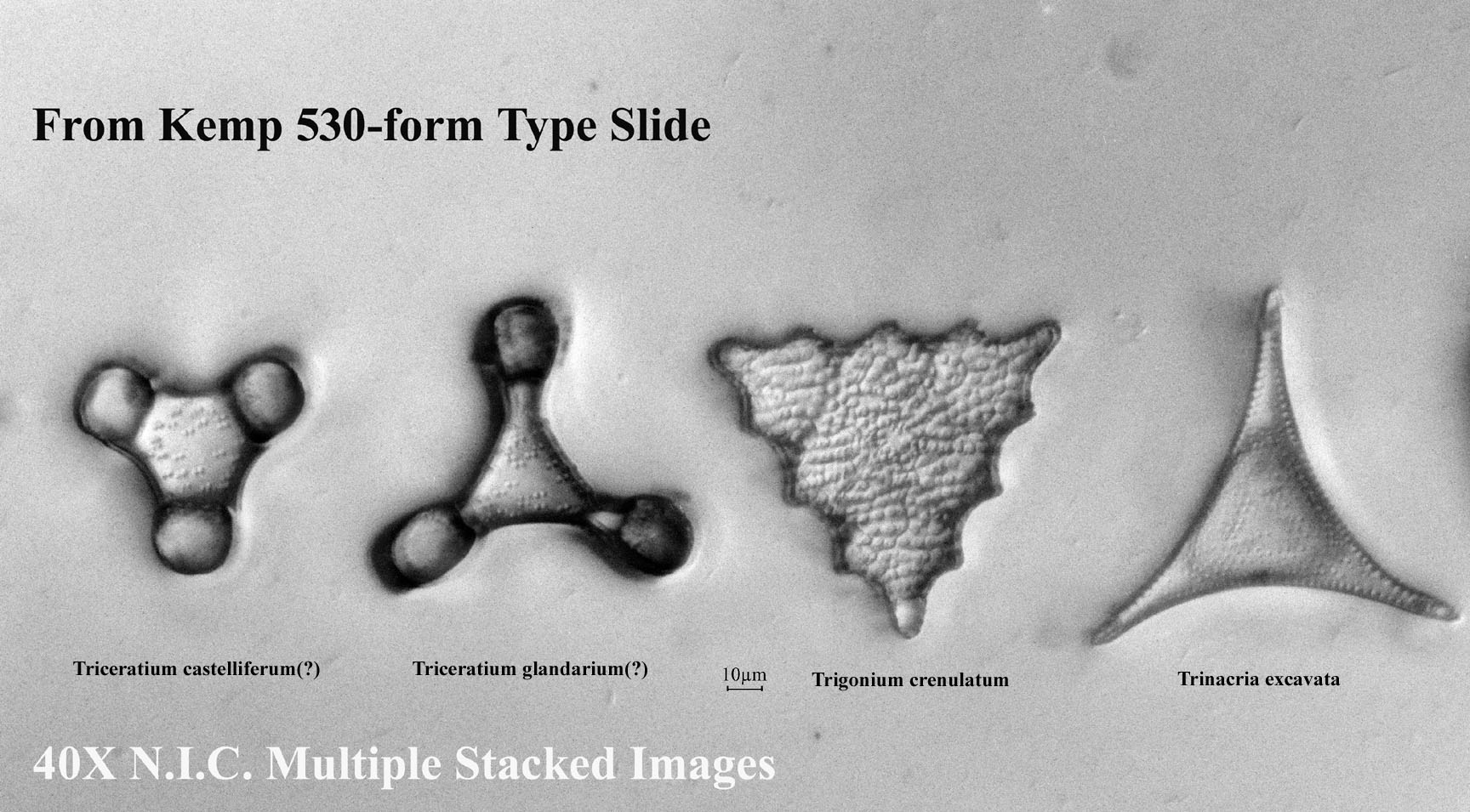
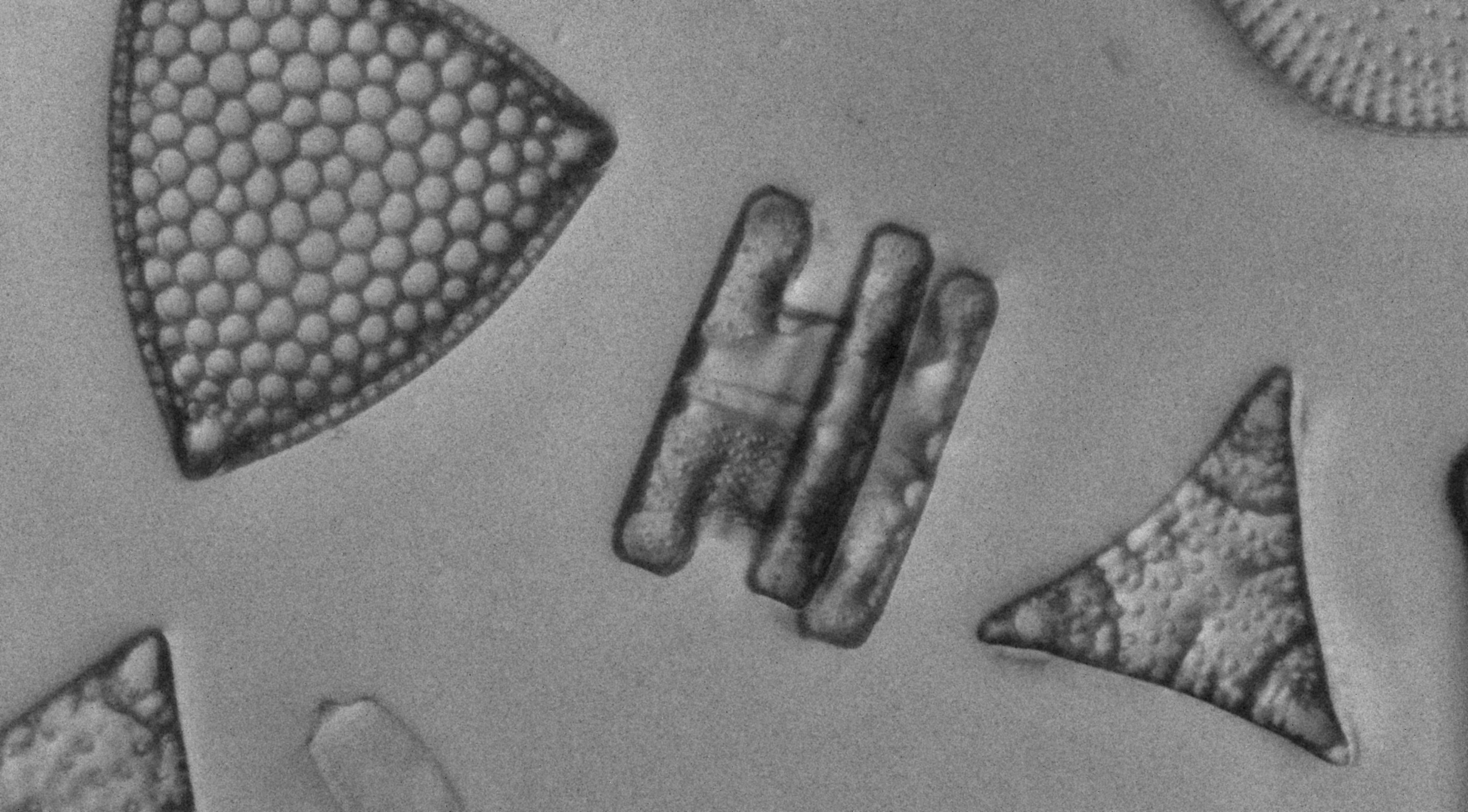
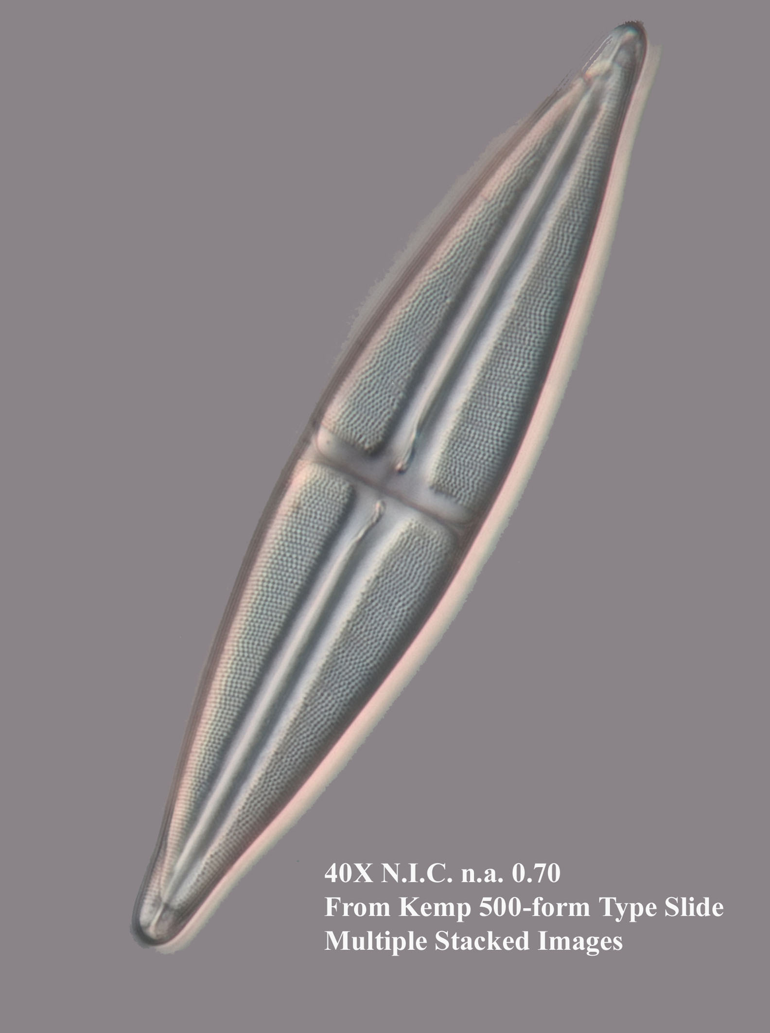
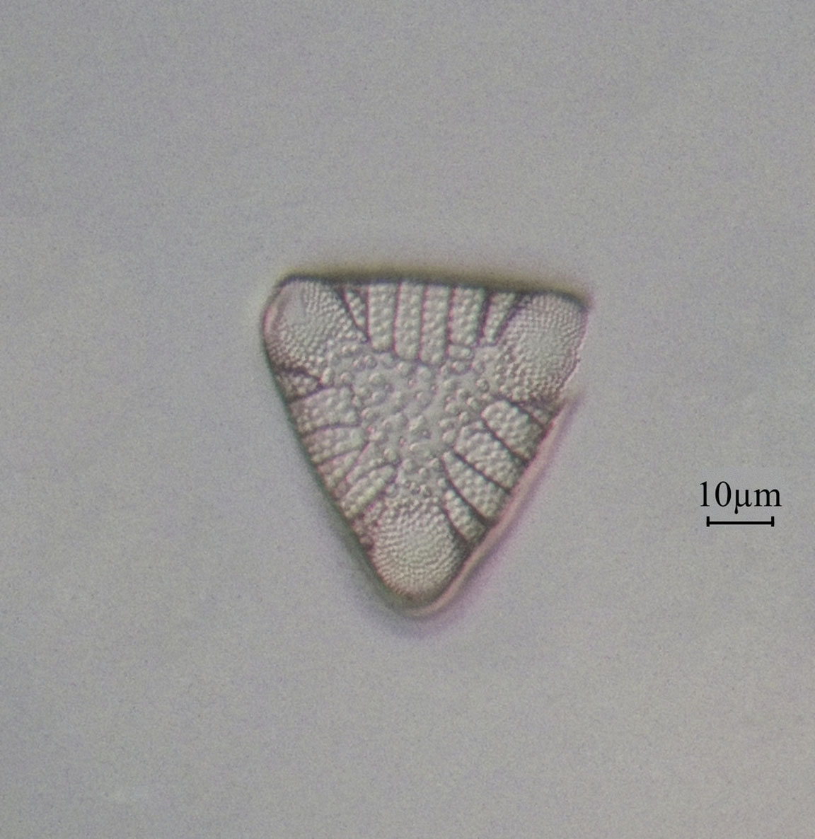
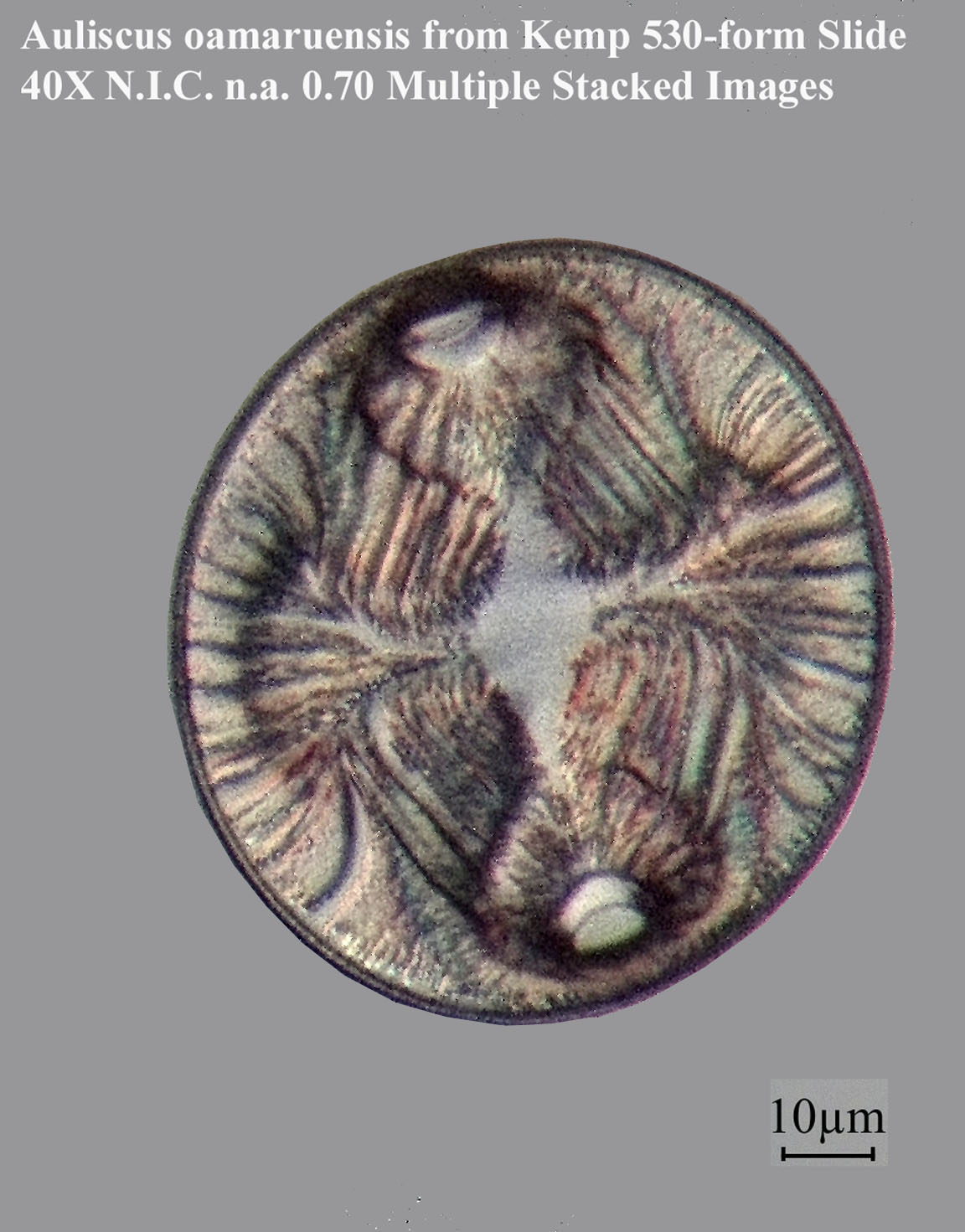
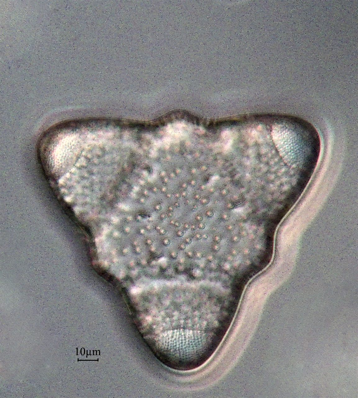
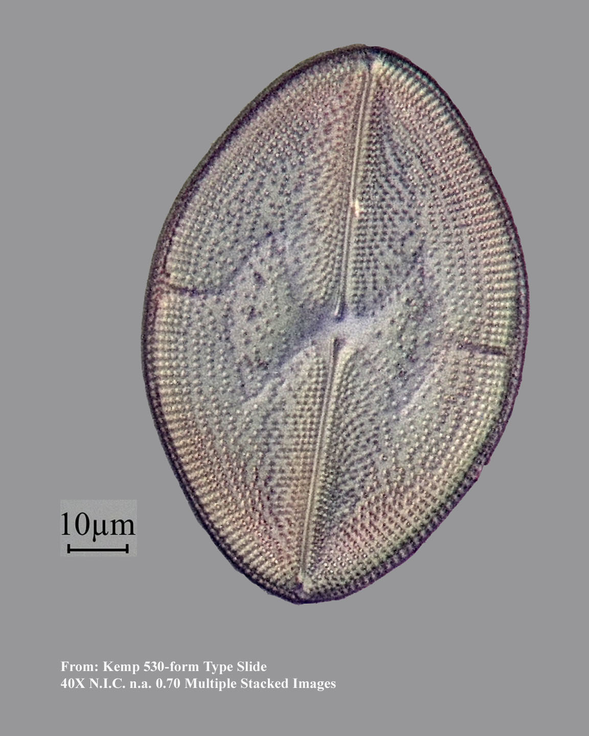
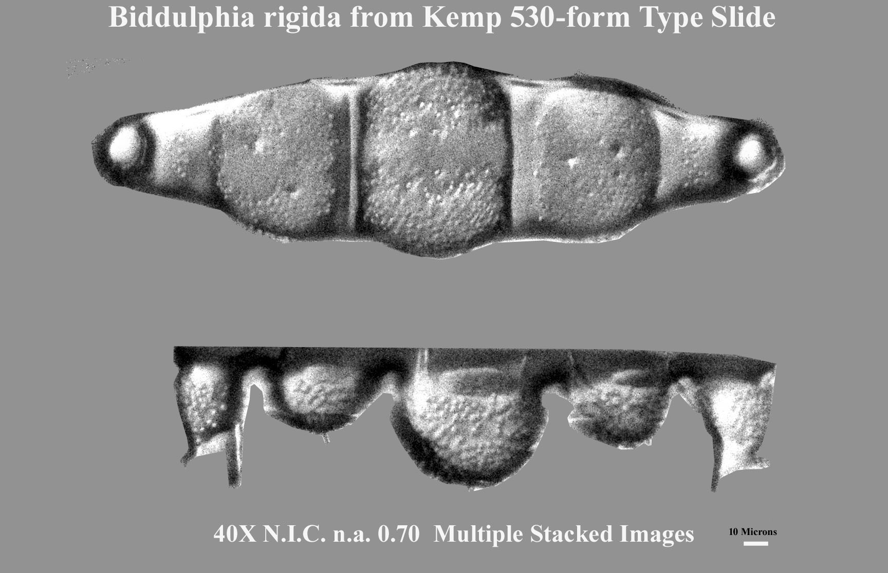
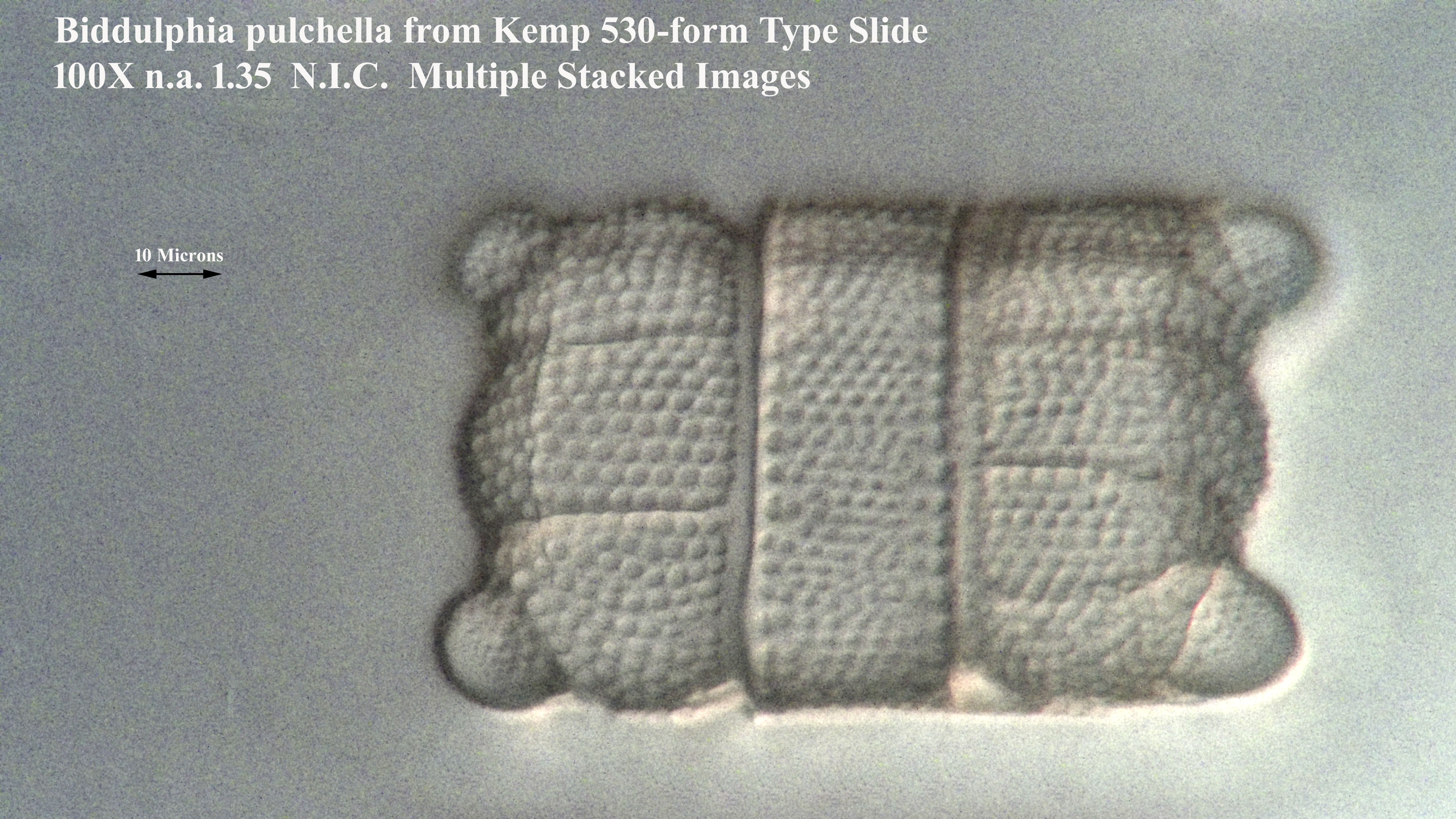
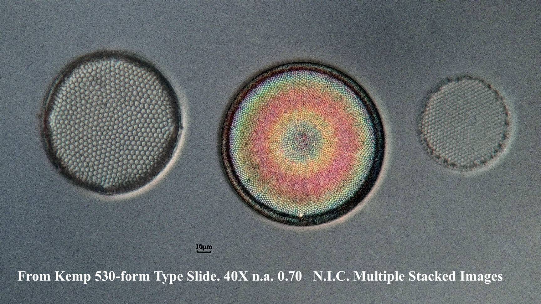
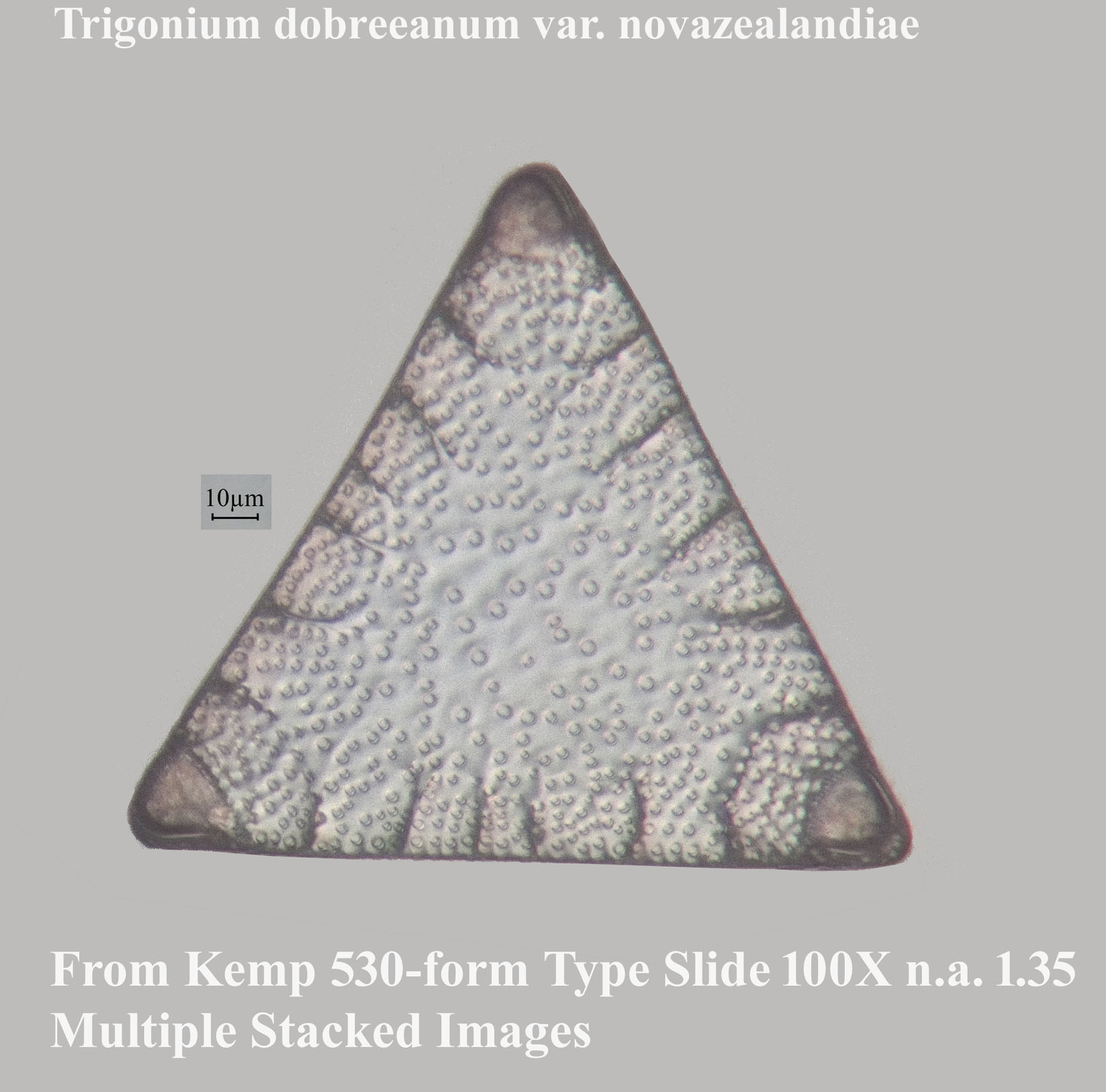
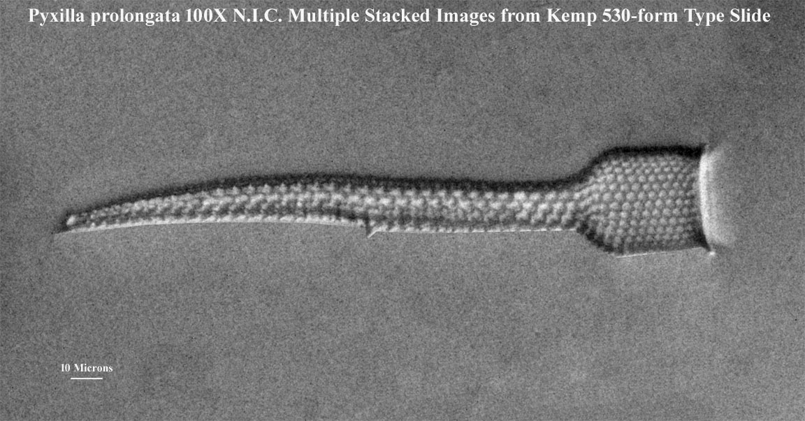
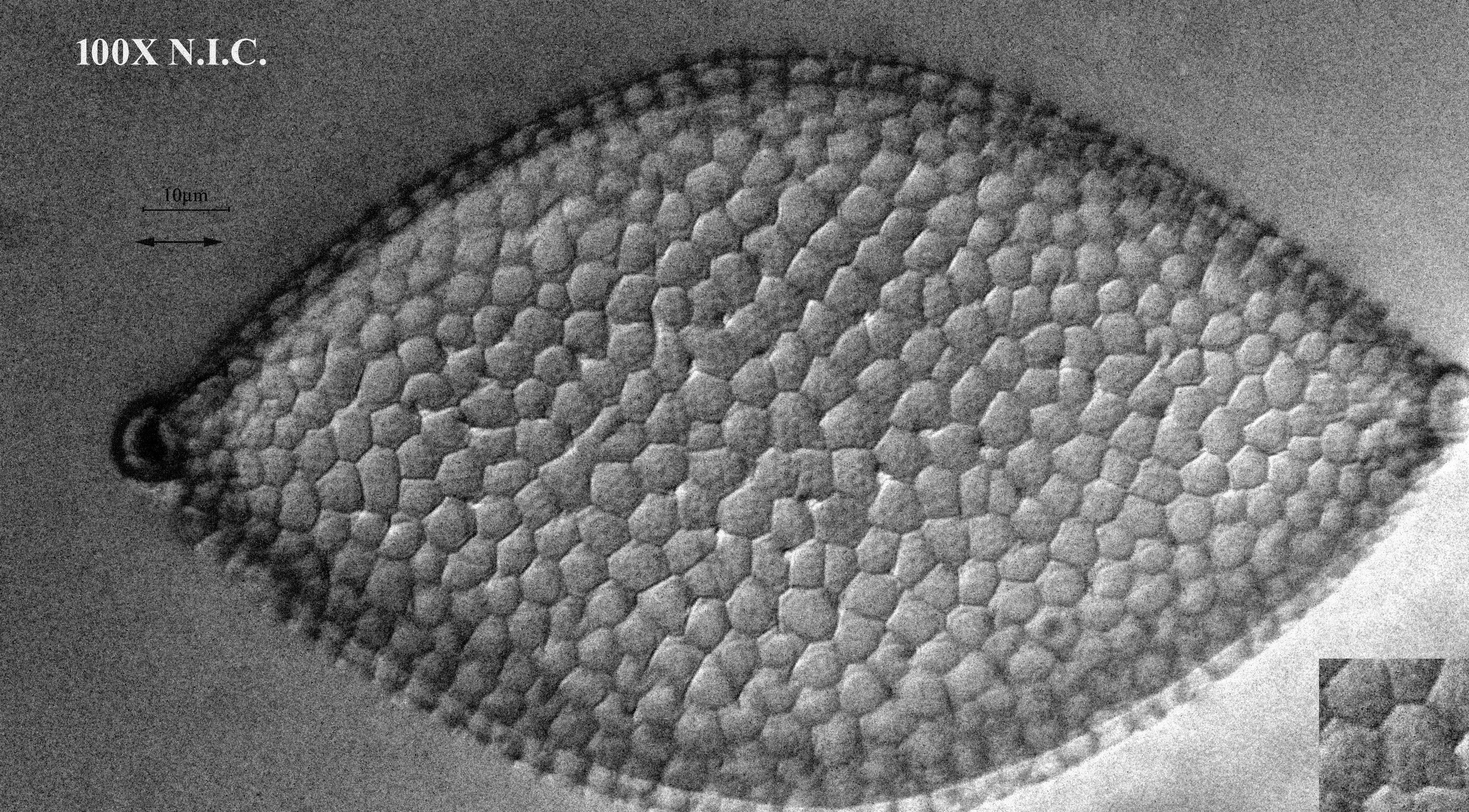
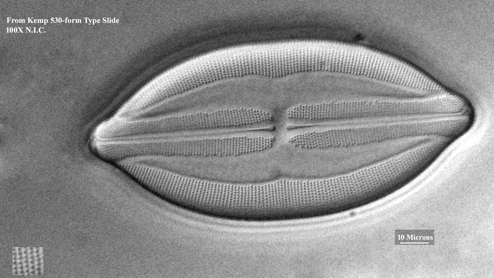
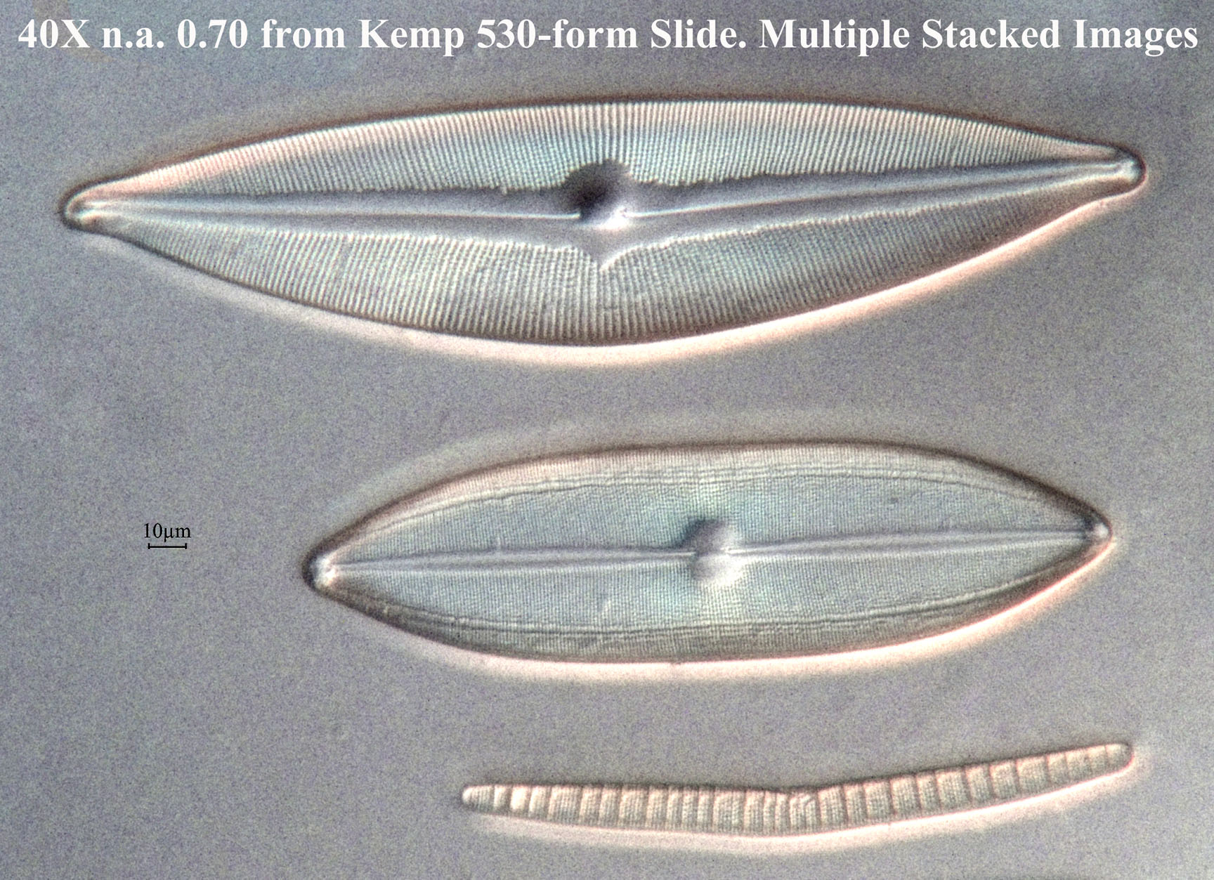
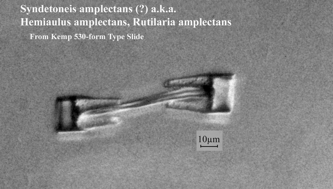
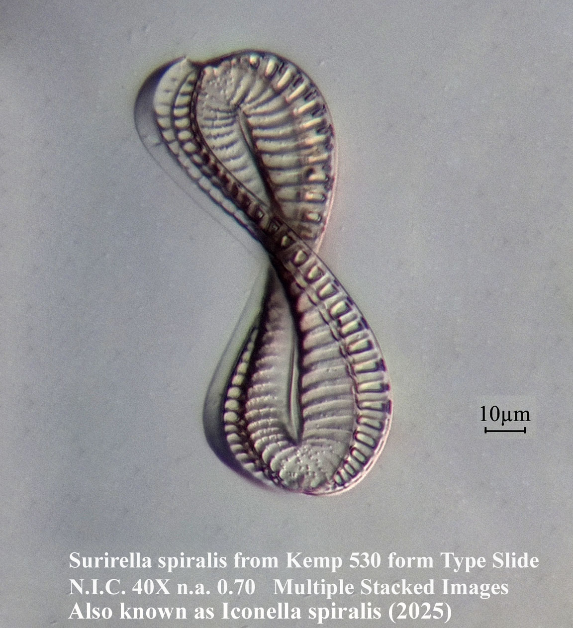
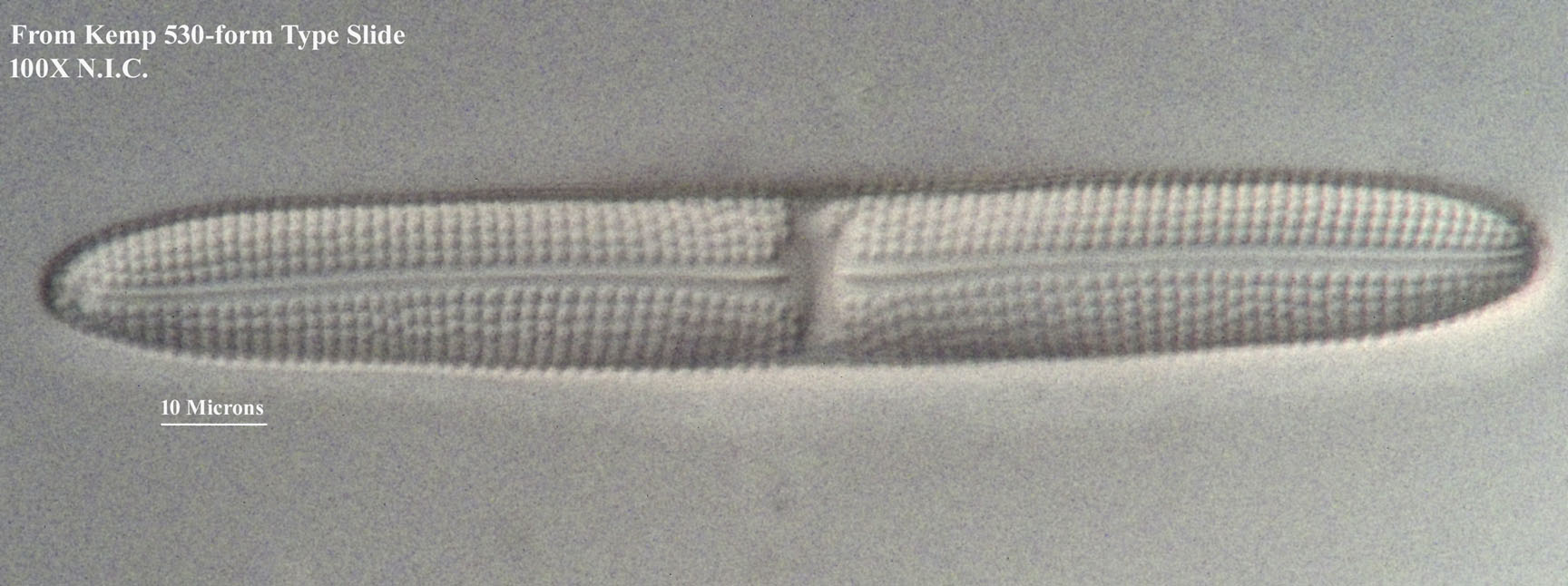
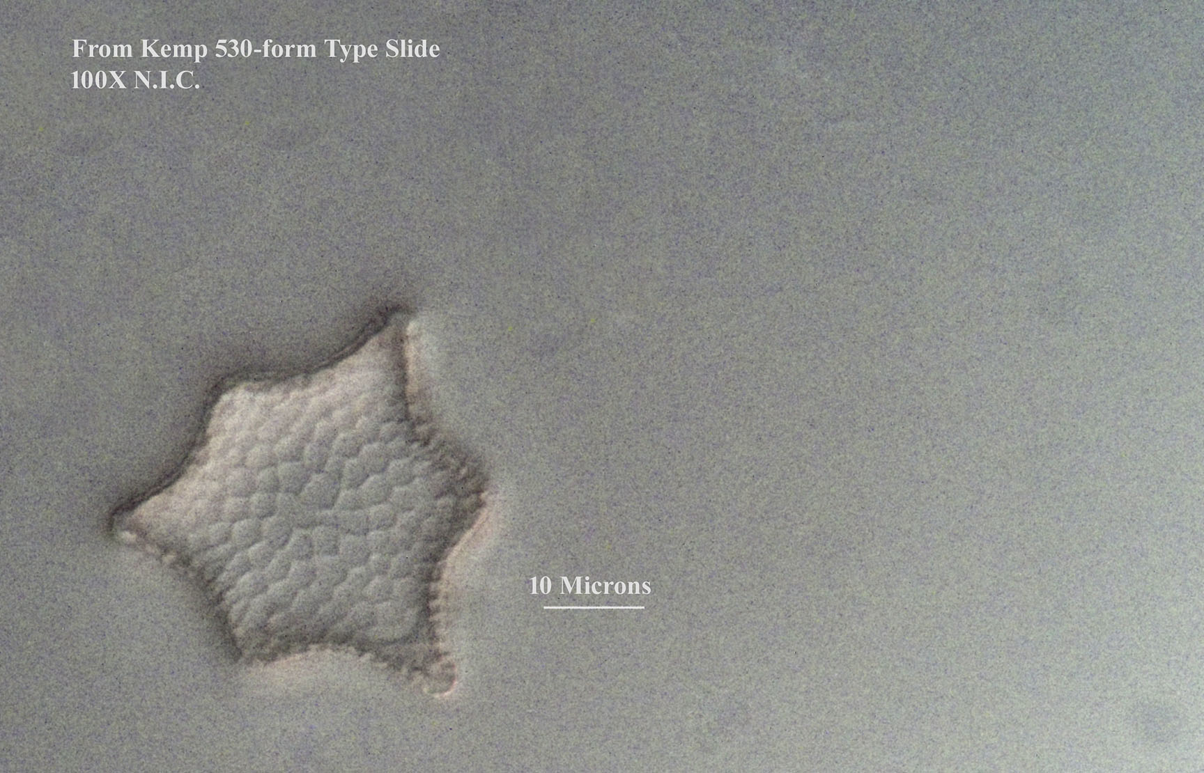
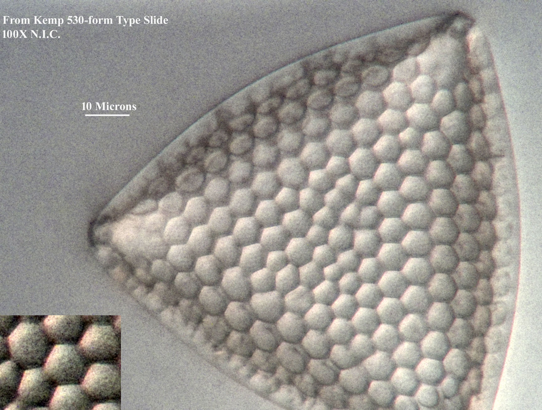
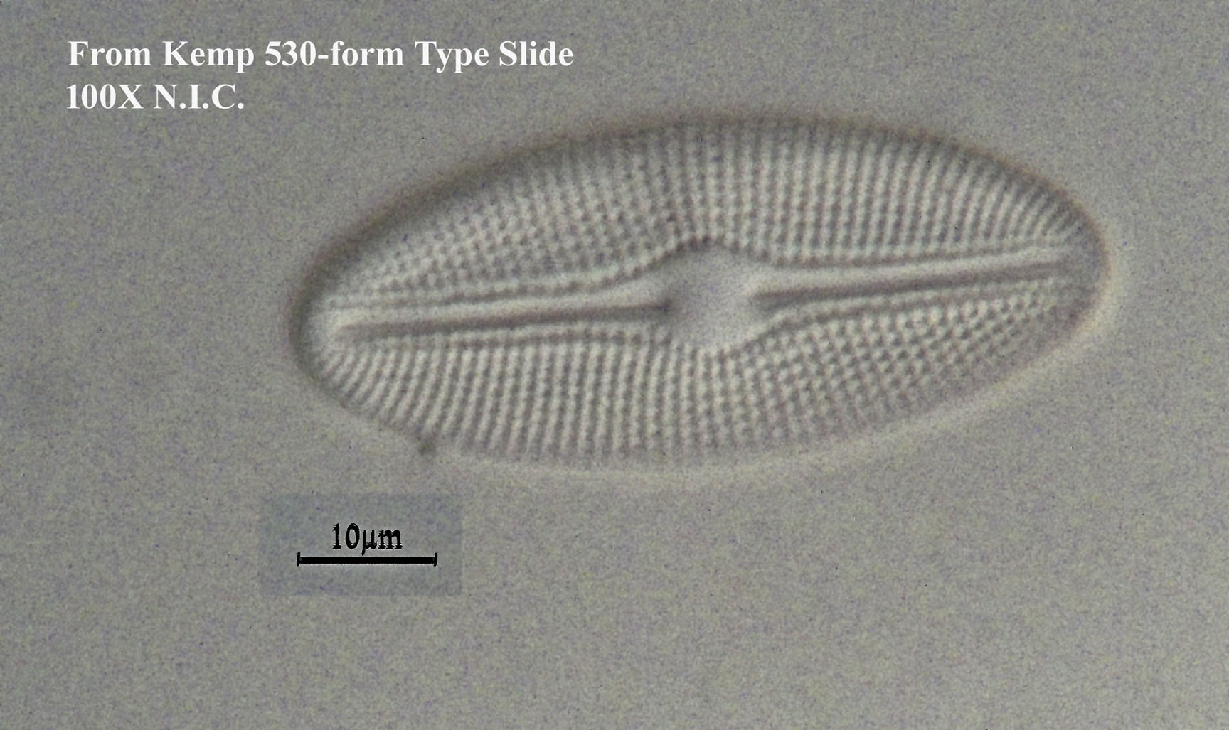
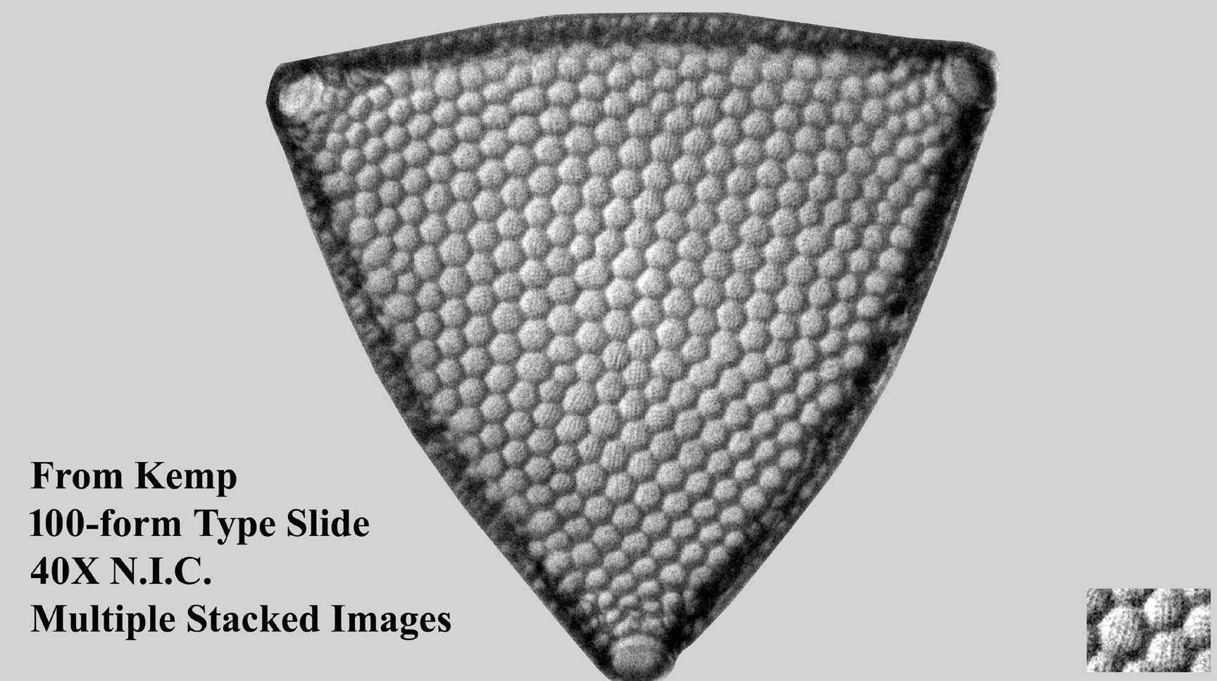
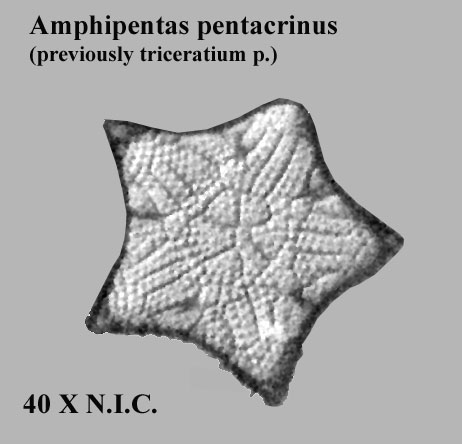
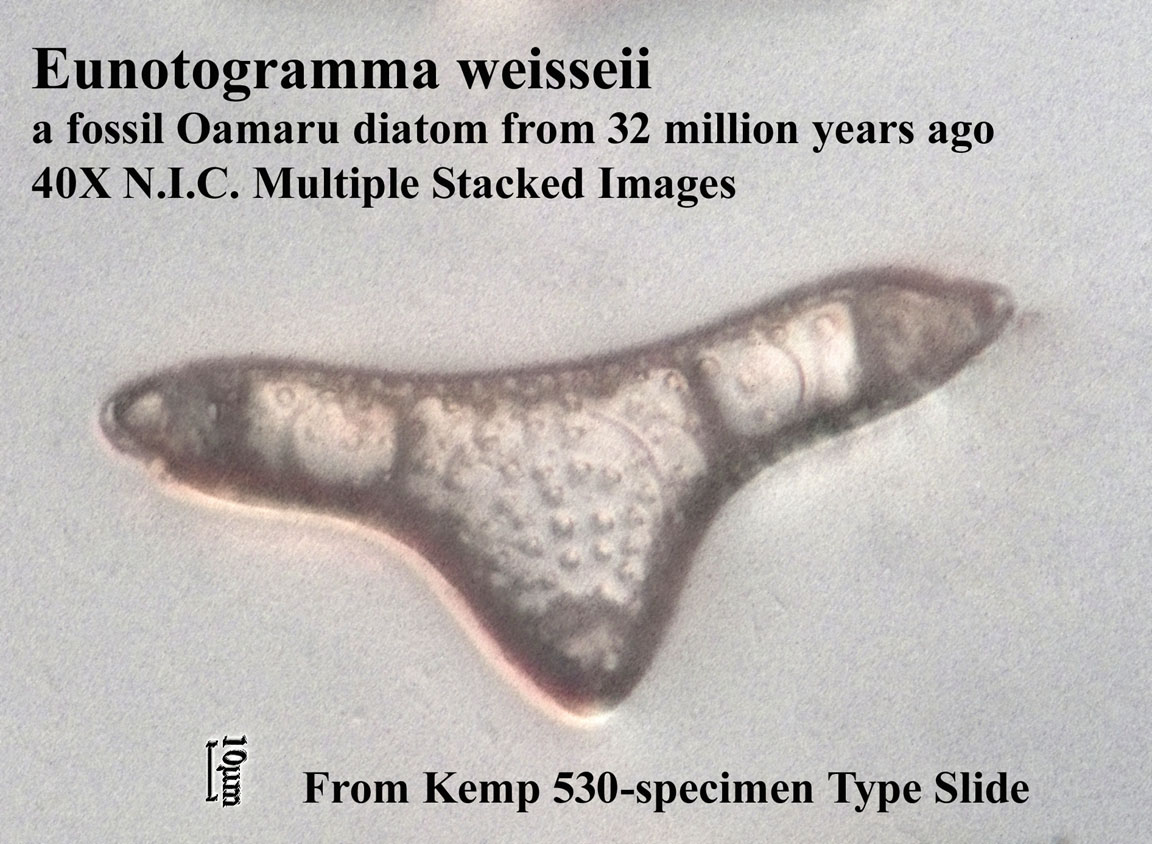
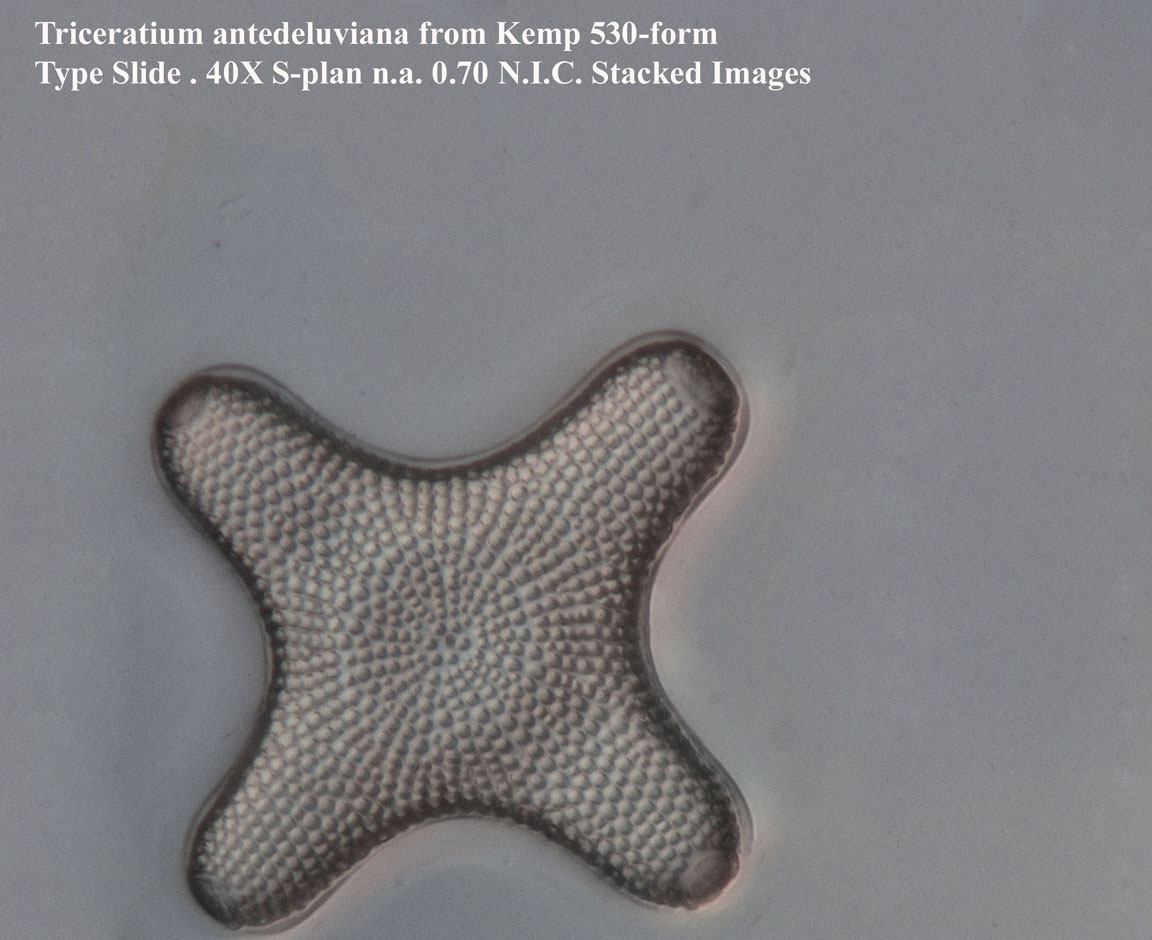
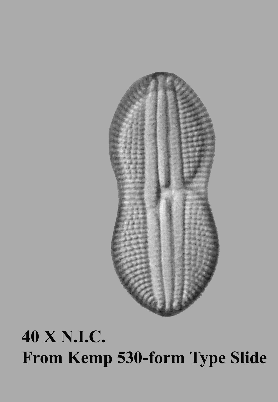
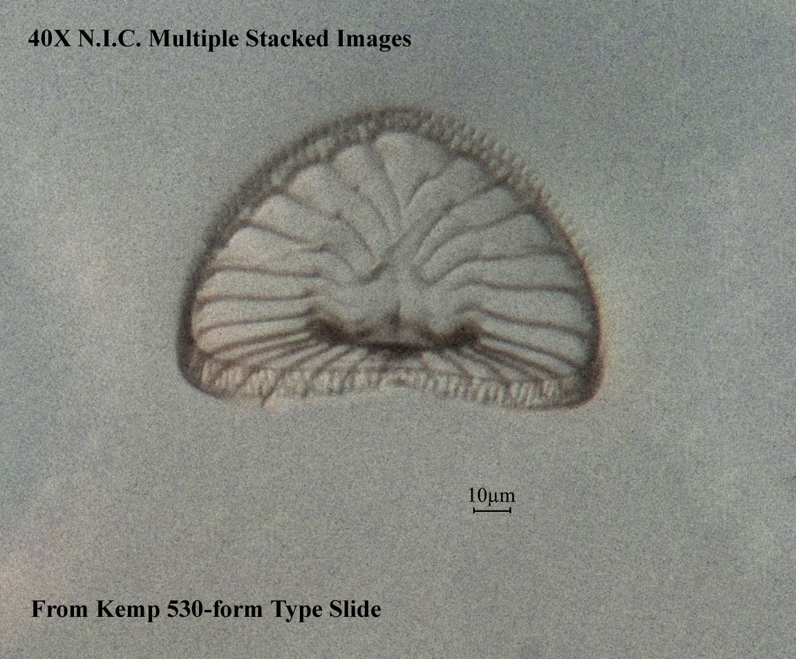
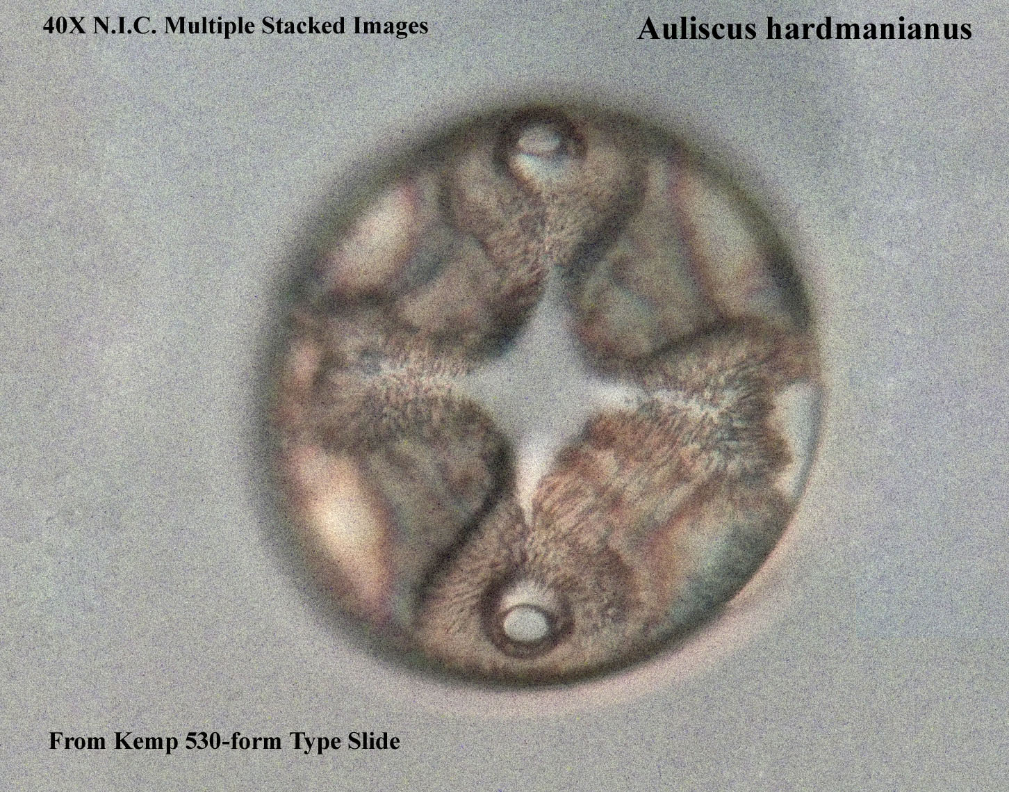
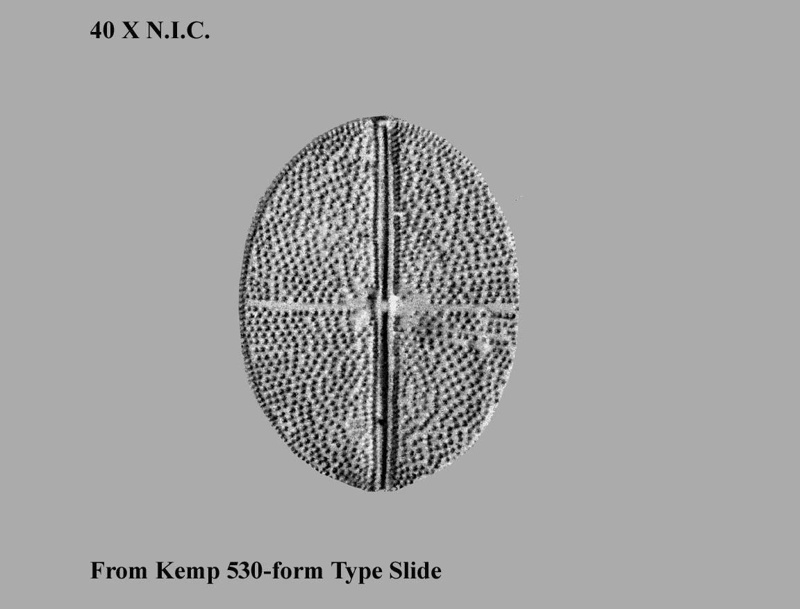
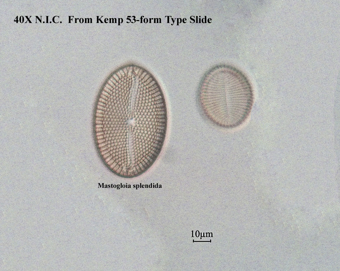
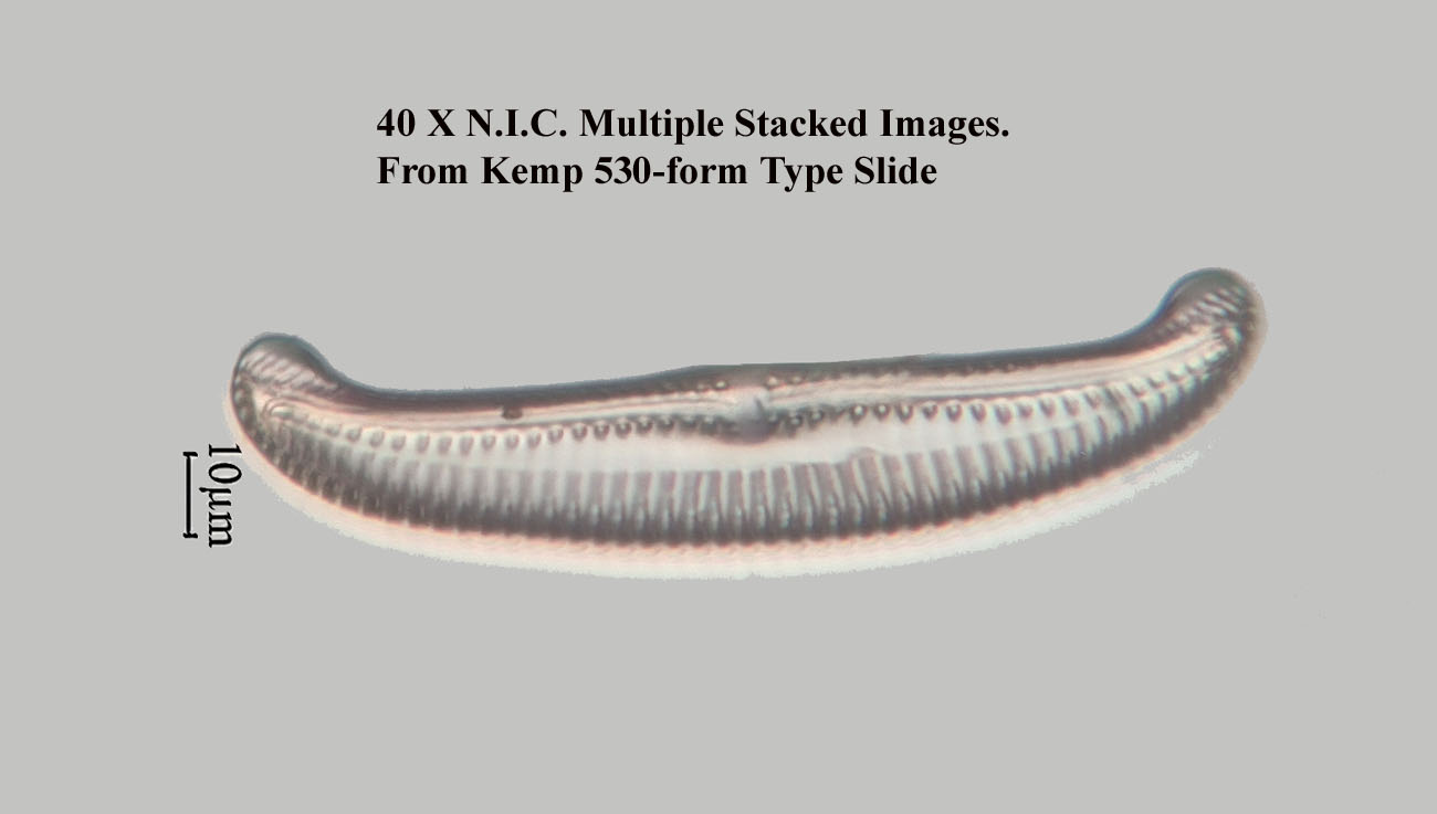
This type slide of 530 forms of diatoms is shown to the left. Above is a low power dark ground illumination view under the microscope. The slide was produced for the author by the late Klaus Kemp in 2004. Kemp took special requests, and on this occasion I asked him to produce a slide of every diatom he had access to. Every specimen is finely preserved with its features clearly visible with the appropriate resolution and magnification, but in a few cases small pieces are missing or the diatom has some other minor defect. The diatoms are mounted on the slide but the coverslip is not mounted directly on the diatoms, being supported by three little metal supports. Although this avoided deforming or breaking the diatoms, it means some diatoms are not as close to the coverslip as they might be otherwise. Unfortunately, he was unable to supply me with a matching species list. Shown below are some of the individual forms shown at higher resolution and magnification via DIC illumination. In 2025 I added some higher quality images and videos illustrating three dimensional structure. Most of the pictures are taken with N.I.C. illumination and are the result of multiple stacked images using Helicon Focus Software.