MICROSCOPE ACCESSORY CASE
WITH POLARIZATION ACCESSORIES AND AN UNUSUAL ACCESSORY STAGE
c. 1910.
DESCRIPTION
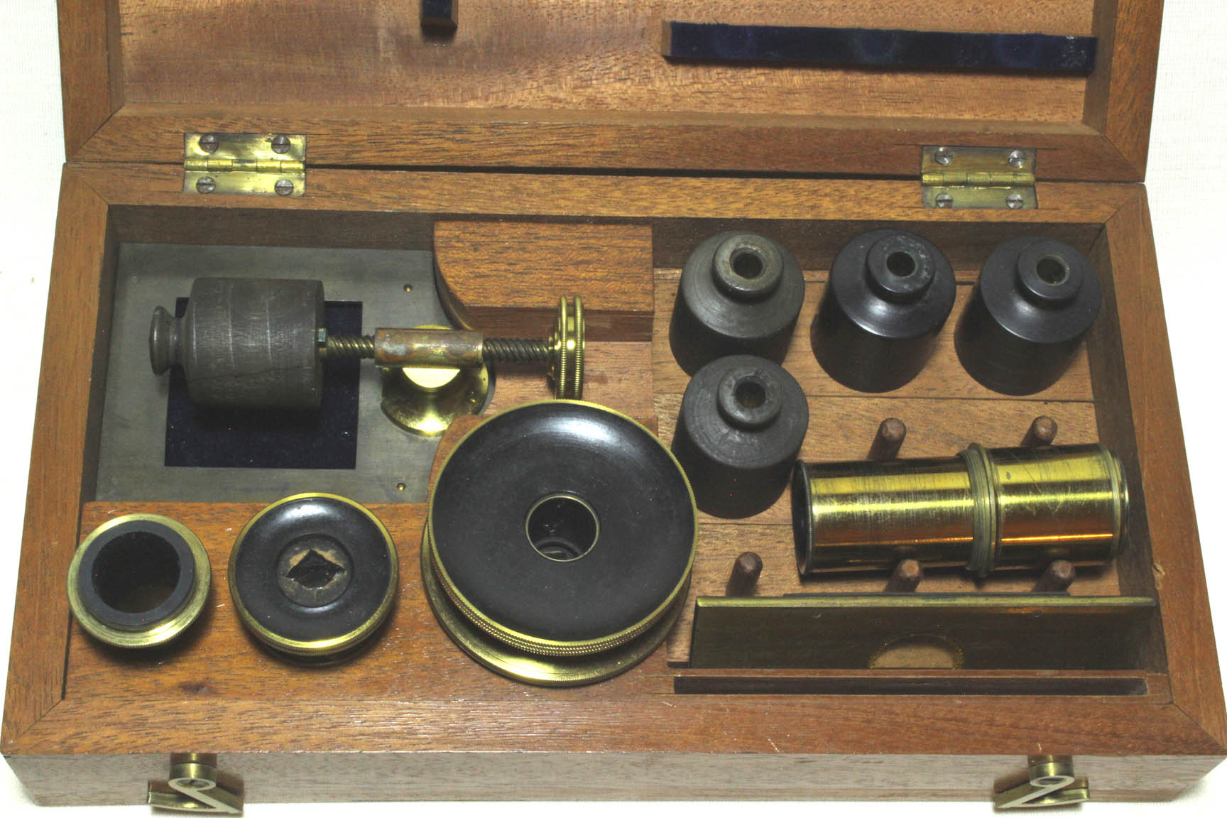
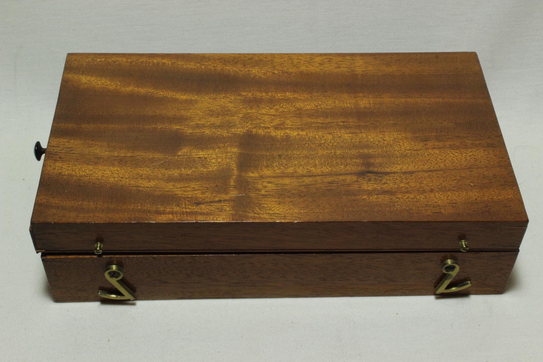
THE CASE:
The case of this kit is made of lightly stained mahogany. The felt on the furniture inside the lid is reddish brown in color. The brass clasps for holding it closed are made of thick brass. The single tiny knub for pulling it out of a larger microscope case is made from an early plastic-like material such as bakelite, which was invented during the first decade of the twentieth century. The characteristics of this case, along with a note found with it dated May 1910, suggests this case is from about 1910.
POLARIZATION APPARATUS:
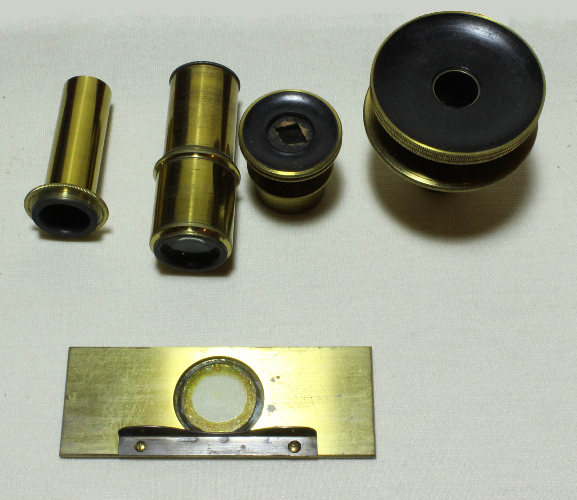 Like other polarization kits meant for qualitative work, this kit comes with an analyzer, a polarizer, and a selenite stage.
The polarizer is attached under the stage or into the substage. The analyzer is fitted above the stage. This kit provides flexibility in the location of the analyzer, which can be used in the eyepiece cap, or removed from the cap and placed in the accessory tube which fits into the end of the draw tube.
Like other polarization kits meant for qualitative work, this kit comes with an analyzer, a polarizer, and a selenite stage.
The polarizer is attached under the stage or into the substage. The analyzer is fitted above the stage. This kit provides flexibility in the location of the analyzer, which can be used in the eyepiece cap, or removed from the cap and placed in the accessory tube which fits into the end of the draw tube.
According to the handwritten instruction with this kit, using the eyecap location for the analyzer reduces or cuts-off part of the field of view, but has the advantage, of giving a better 'defining power.' Use of the eyecap position therefore was recommended for determining the 'form and shape of crystals and other objects... and obtaining a sharper outline.' The in-tube position was otherwise generally recommended. Two sets of handwritten instructions came with the case. One is 'W. Smith's directions' and the other is signed by 'W. Walton.' Walton's was meant to be a supplement to the instructions by Smith.
ACCESSORY STAGE:
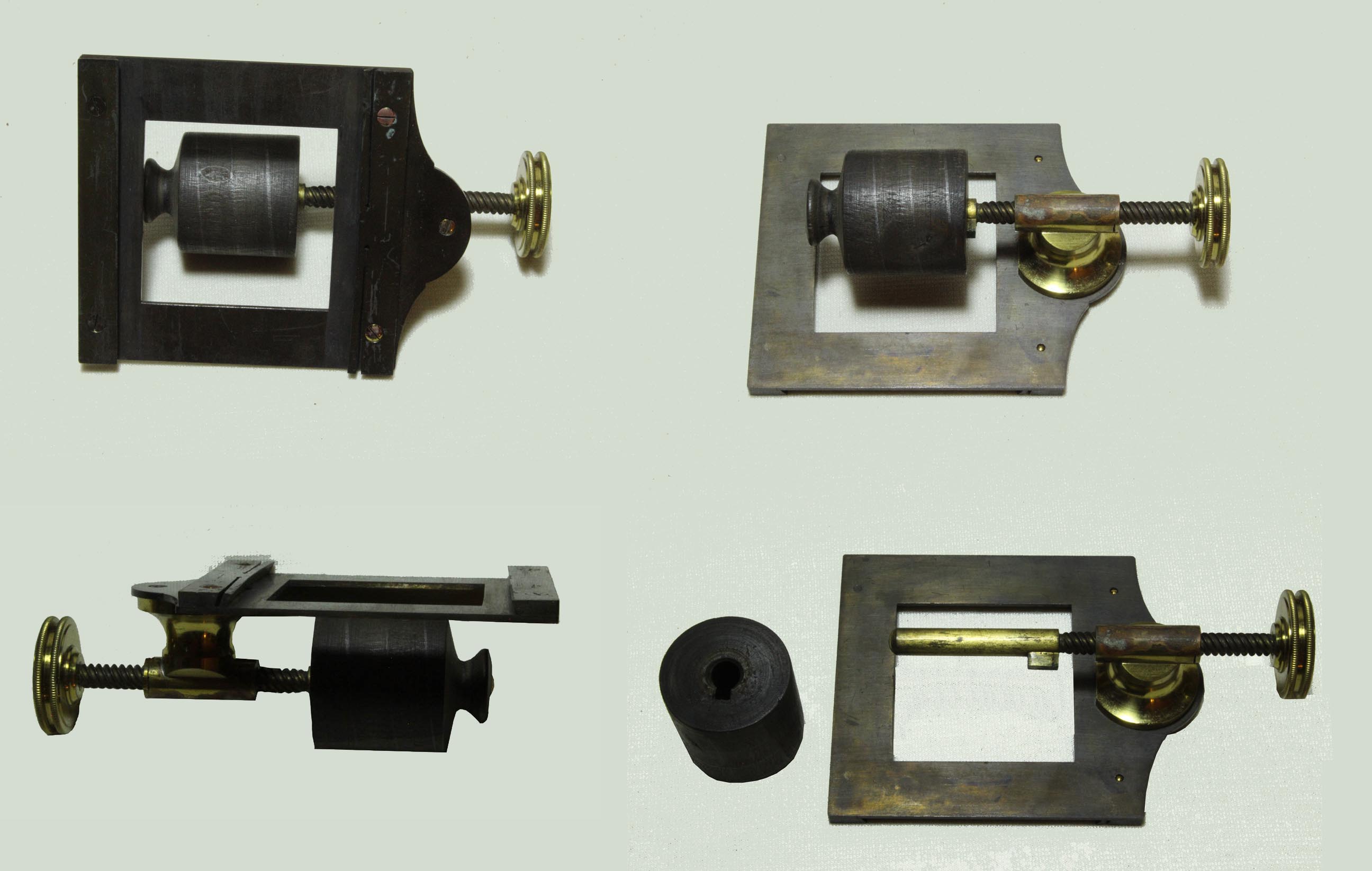 This stage is a bit of a mystery. Its exact purpose to date has eluded the author and many of his friends familiar with both old and new microscopes. It consists of a support which allows a wooden spool to be attached and then rotated. Inscribed on each spool is a line drawn in pencil. As the rotation of the knurled knob advances the spool, the pencil line is passed through exactly the same point. In other words, as the spool rotates, whatever subject is attached following this line, would be successively brought under the same point in the field of view. This suggests the possible use of this stage for examining something like thread. twine, rope or perhaps fabric.
A more remote possibility is that the spools were to be used to hold a large number of three dimensional objects such as crystals, cemented along the line. Whatever is its true intended purpose, I have been unable to find any reference to this in the literature. It may be that this is a unique item, made by special order. It would not seem to have anything to do with the polarization set, as the latter would be used with transparent objects, unless crystals were to be mounted along the lines, but if this were the case this would seem to be a poor method of mounting them.
This stage is a bit of a mystery. Its exact purpose to date has eluded the author and many of his friends familiar with both old and new microscopes. It consists of a support which allows a wooden spool to be attached and then rotated. Inscribed on each spool is a line drawn in pencil. As the rotation of the knurled knob advances the spool, the pencil line is passed through exactly the same point. In other words, as the spool rotates, whatever subject is attached following this line, would be successively brought under the same point in the field of view. This suggests the possible use of this stage for examining something like thread. twine, rope or perhaps fabric.
A more remote possibility is that the spools were to be used to hold a large number of three dimensional objects such as crystals, cemented along the line. Whatever is its true intended purpose, I have been unable to find any reference to this in the literature. It may be that this is a unique item, made by special order. It would not seem to have anything to do with the polarization set, as the latter would be used with transparent objects, unless crystals were to be mounted along the lines, but if this were the case this would seem to be a poor method of mounting them.
HISTORY OF POLARIZATION WITH THE MICROSCOPE
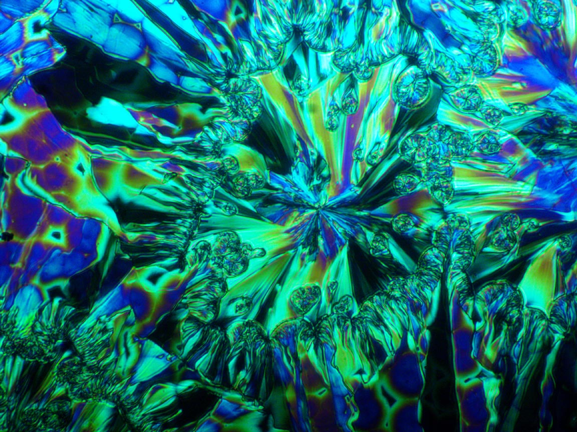
Photomicrograph of a Slide of Salicine Taken with a Selenite and Crossed Polars.
The polarizer and analyzer with this set make use of Nicol prisms. These were invented by William Nicol, a lecturer at the University of Edinburgh in the early 19th century. They were made by splitting a piece of Iceland spar, or calcite and then cementing the halves together with Canada balsam. This separates the light rays into 'ordinary rays' which are reflected back out of the prism (or absorbed by the blackened side of the prism), and the 'extraordinary rays' which pass through the prism linearly polarized. The first use of this in a microscope was credited to Talbot in 1834.
Certain materials exhibit beautiful patterns when viewed between 'crossed polars.' )
Crossed polars which means that the orientation of the polarizer and analyzer are at 90 degree angles to each other; if no subject were placed between the polarizer and analyzer, the field would be dark. When an anisotropic material is placed between polarizer and analyzer, an image, altered by the properties of the subject appears, often with colors and shapes that are both informative and beautiful. A further change can be imparted by the use of a wave plate (also known as a compensator, or retardation plate), between the polarize and analyzer, either above or below the object being studied. The most common material used for this purpose until the 20th century was the mineral selenite. Selenite often imparts additional colors to the image. An otherwise clear or bland subject may appear spectacular when viewed with crossed polars with or without a wave plate, and often, though not always the wave plate adds even more appeal. This effect can occur not only with minerals but also certain chemicals, and even things like bone or insect parts.
In the nineteenth and early twentieth century the commonest use for polarized light microscopy was for entertainment, but serious geological work could be done with a petrographic microscope which can allow precise identification of a variety of crystals and rocks. This more serious work requires much more than is provided in the kit supplied here. For more information about petrographic microscopes and especially their history,see: Kile, Daniel: The Petrographic Microscope. Supplement to The Mineralogical Record November-December 2003. A petrographic microscope by Leitz from about 1940 is shown on this web site.