'LENTICULAR MICROSCOPE'
MAKER: MORITZ PILLISCHER
c. 1850
Serial No: 153
Please Click On Any Picture for a Larger Version
DESCRIPTION: The trough-like rectangular stage of this microscope is made of oxidized brass and is about 0.9 mm thick. The inside dimensions of the trough are about 27.5 x 77 mm which clearly reveal it was designed to be used with standard 1 x 3 inch (25.5 X 77 mm) slides. A small black and white disk, 22.5 mm in diameter was also supplied for viewing opaque objects. There is no apparent way for securing this disk on the stage however.
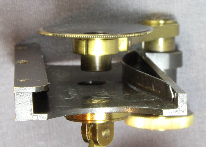 Looking at the short end of the stage, it is easy to see that it has basically a shallow, U-shape. On the short vertical edge on the left is attached a narrow horizontal plate which is used to support a leaf spring that pushes down onto the specimen slide.
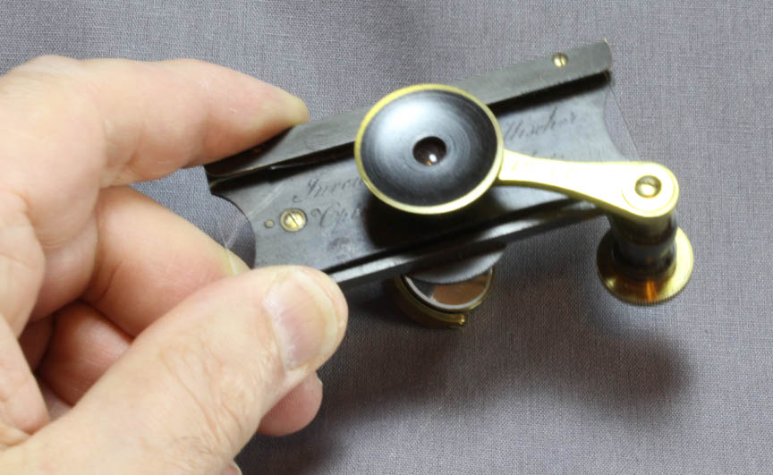
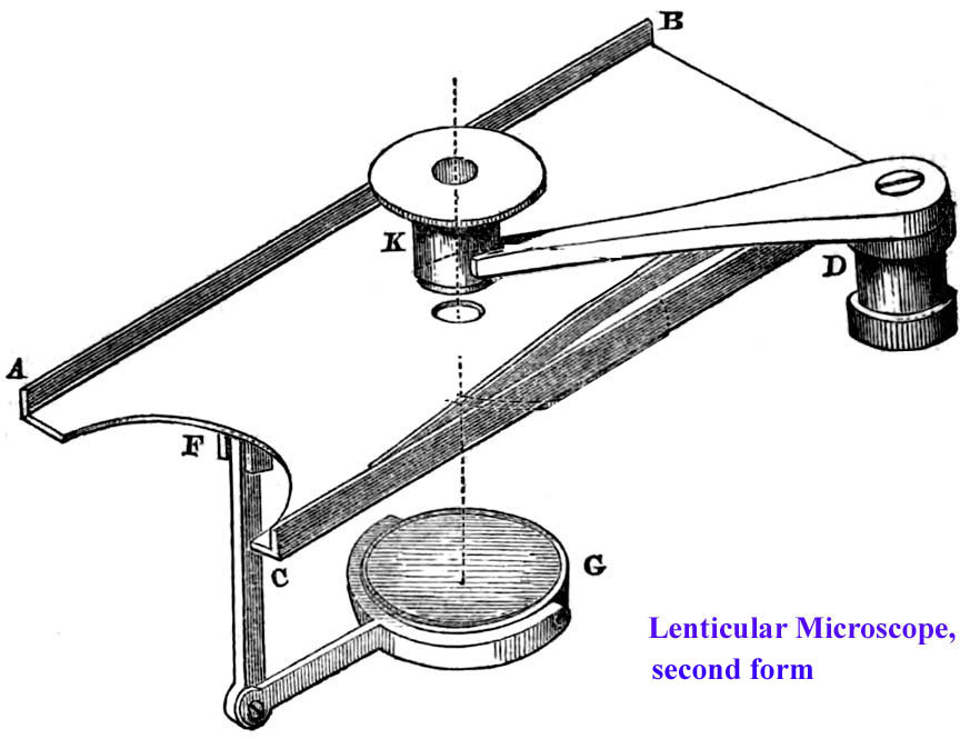
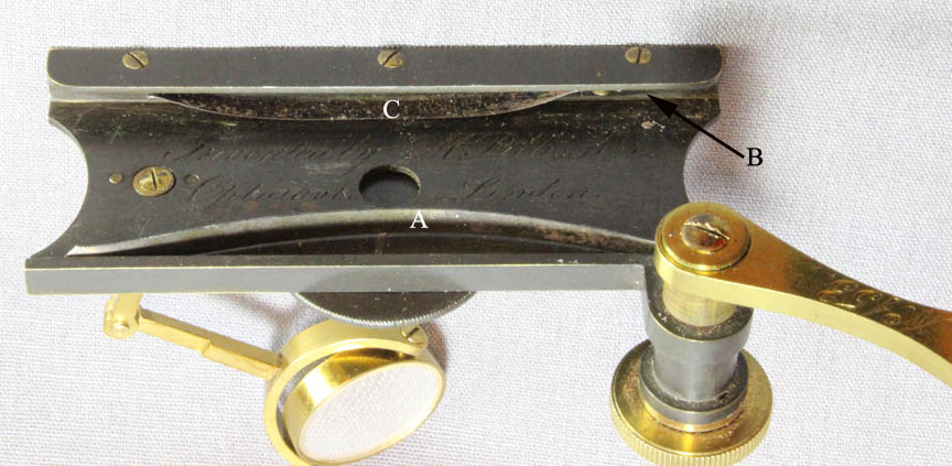 Shown in the illustration to the right, the two bow-shaped steel springs for holding the slide can be seen. One, A, bowing out horizontally, pushes the specimen slide against the straight rear edge, B, of the stage. The other, C, bowing downward, pushes the slide against the surface of the stage. Each spring is held to the stage at one end by a single tiny lacquered brass screw. The pressure of these two springs allows the slide to move smoothly and accurately from side to side as the whole specimen is examined.
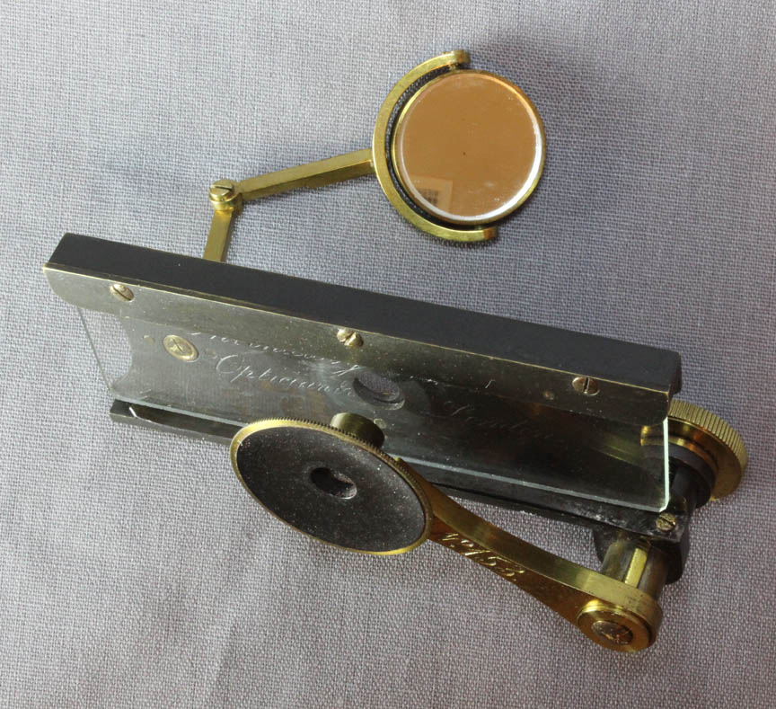
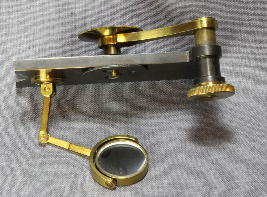
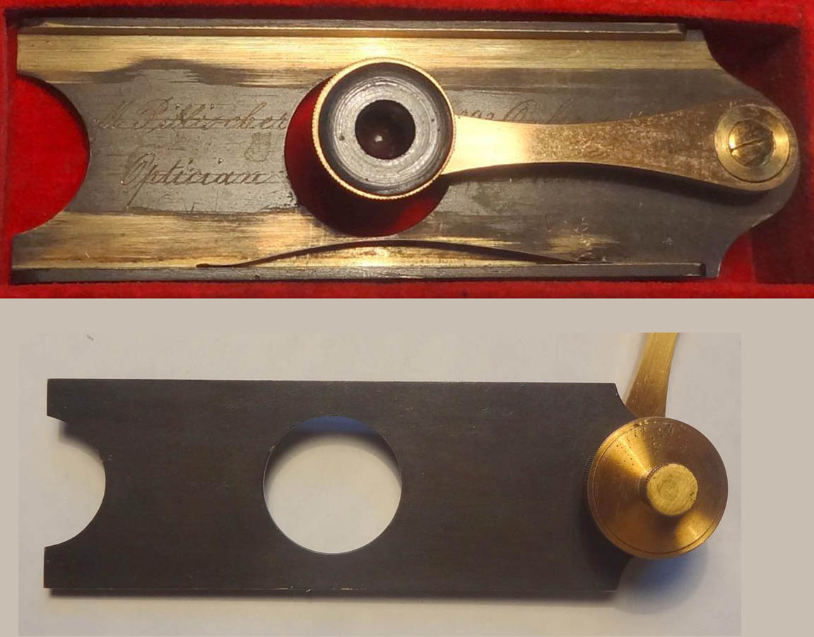
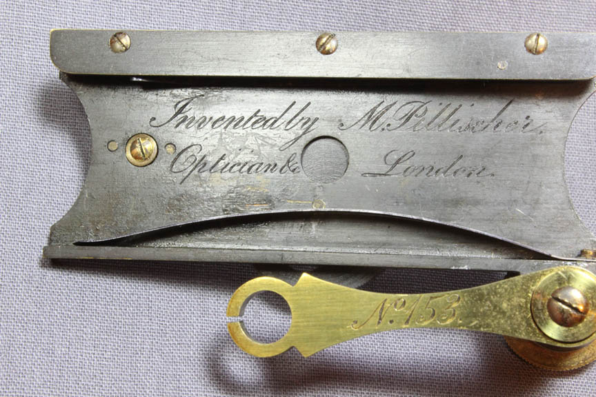 The stage is signed in script 'Invented by M. Pillischer., Optician &c. London.'. The lacquered brass arm is signed in script: 'No. 153'. The arm accommodates any one of the three simple lenses which is pushed into the split ring at the end of the arm.
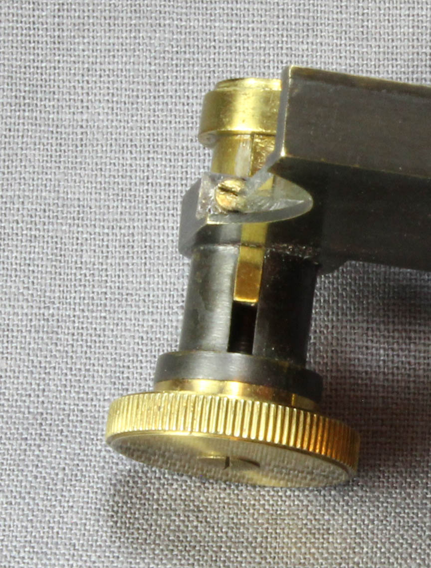 A lacquered brass knob acts on the arm support to raise or lower the arm for focusing. The knob is 'trapped' at the bottom of the focusing assembly so that as it is turned, it remains in place while its attached fine screw raises or lowers the arm support. Because the arm support has a protruding piece, or 'key,' that fits into the sleeve with a groove, there is no rotation imparted to the arm support during focusing. The lens arm itself is held on the support by a screw and washer that allows it to swivel from side to side over the entire specimen area.
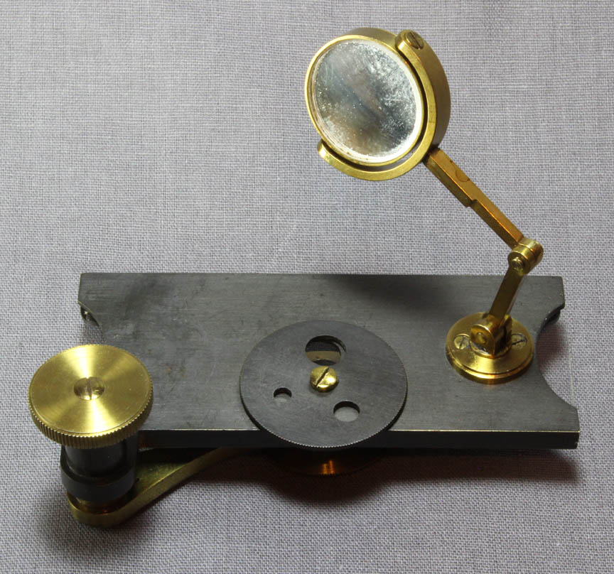 The brass mirror-cell which holds a single concave mirror, can pivot on the end of a double jointed tailpiece. The tailpiece can also rotate in its round lacquered brass support boss where it is attached to the bottom of the stage.
A rotating aperture disk of oxidized (or blackened) brass, is also attached to the underside of the stage to regulate illumination of the specimen. There is no mechanism other than friction to hold the aperture over the stage opening. The three apertures measure about 6.5 mm, 4.5 mm, and 3 mm in diameter, while the stage opening is 7.5 mm in diameter.
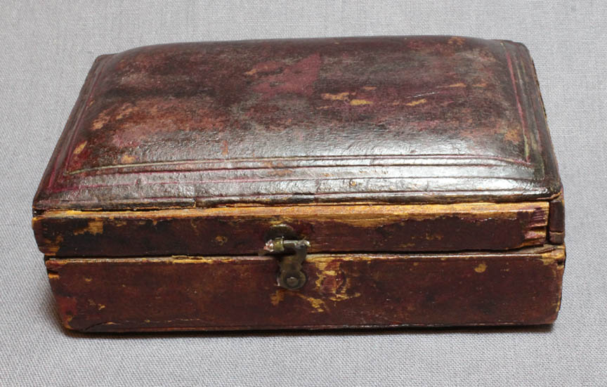
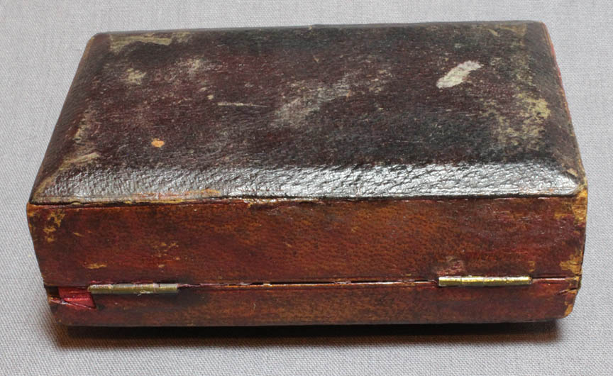
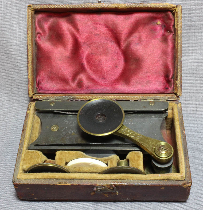
The storage case for the microscope, which measures about 100 mm X 58 mm and is about 37.5 mm thick, is a thick rectangular shape with a domed lid. It is covered with red leather on the top and sides, and black leather on the bottom. The inside of the case is lined in red silk on the lid and red velvet for the rest. The red color has faded from the velvet, which is now partly a tan color.
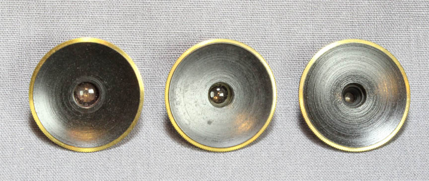
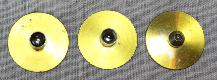
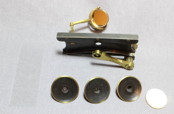 Accessories include a black and white ivory disk, and a total of three lenses, each with an outside diameter of about 26 mm. Two of the lenses are stored in slots in the case while the third remains mounted in the arm of the microscope.
The original microscope may have included a glass depression slide as one is mentioned in early accounts of this microscope and is found with other surviving examples. A replacement depression slide has been added to this outfit and, like the original, is stored on the stage.
Accessories include a black and white ivory disk, and a total of three lenses, each with an outside diameter of about 26 mm. Two of the lenses are stored in slots in the case while the third remains mounted in the arm of the microscope.
The original microscope may have included a glass depression slide as one is mentioned in early accounts of this microscope and is found with other surviving examples. A replacement depression slide has been added to this outfit and, like the original, is stored on the stage.
Dr Joseph Zeligs has been very helpful in analyzing the construction of the lenses accompanying this microscope, and has concluded they are all of the Coddington type. Although X-ray images of the high power lens suggested it might be a doublet, careful analysis of the lens, including inspection with a stereo microscope, showed that it too is a single lens, and most likely a Coddington, and occupies only a small part of the housing nearest the specimen. It is notable that under the stereo microscope, the curved surfaces of the high power lens are quite pitted in places, i.e. it is not a perfectly polished lens.
HISTORY OF THE MORITZ PILLISCHER LENTICULAR MICROSCOPE (written with the very kind collaboration of Dr Joseph Zeligs)
 Apparently the first form of Pillischer's Lenticular microscope had only a stage with a central hole, and the arm to carry the lenses. No substage mirror or aperture wheel was present as is illustrated in this early example. I am not aware of any contemporary illustration of this first model in any publicaion of the day. Notice the side supporting the arm curves outward, convex, while the other side curves inward and is concave. Pillischer must have quickly realized that if this microscope was to be used with liquids, it would need a substage mirror to allow it to be illuminated, yet remain horizontal.
 As shown here, and also illustrated by Allan Wissner's example, serial number 32, the second version of Moritz Pillischer's Lenticular Microscope was made initially
with no wheel of apertures and the stage was a simple U-shape in profile. There was only one bow spring to hold the slide against the opposite long side of the stage. Since there was no spring to hold the slide against the stage bottom, the slide could easily pop up. In that second form, the short side of the stage near the focusing knob was straight across
while on the opposite side there was a half-round cutout to allow the slide to be easily grasped by one's fingers and moved side to side. The signature on these early versions, including the serial number, was engraved on the stage; nothing was engraved on the lens arm.
As shown here, and also illustrated by Allan Wissner's example, serial number 32, the second version of Moritz Pillischer's Lenticular Microscope was made initially
with no wheel of apertures and the stage was a simple U-shape in profile. There was only one bow spring to hold the slide against the opposite long side of the stage. Since there was no spring to hold the slide against the stage bottom, the slide could easily pop up. In that second form, the short side of the stage near the focusing knob was straight across
while on the opposite side there was a half-round cutout to allow the slide to be easily grasped by one's fingers and moved side to side. The signature on these early versions, including the serial number, was engraved on the stage; nothing was engraved on the lens arm.
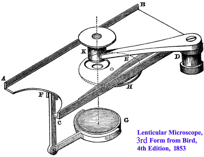 As illustrated by serial number 391, the third form had the understage wheel of apertures added, but was otherwise the same. It is this third form that Golding Bird showed in the fourth edition of his book in 1853 (see below).
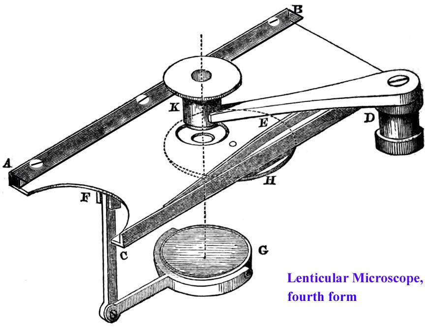 Starting no later than number 45, a horizontal strip, AB, was attached to the top of the long rear edge of the stage. A second leaf spring was attached to its underside to press down on the specimen slide. Now the specimen slide was held securely by two bow springs. The signature remained on the stage. The design of the case changed and
it became like the one for the microscope being described as illustrated at the top of this page.
Starting no later than number 45, a horizontal strip, AB, was attached to the top of the long rear edge of the stage. A second leaf spring was attached to its underside to press down on the specimen slide. Now the specimen slide was held securely by two bow springs. The signature remained on the stage. The design of the case changed and
it became like the one for the microscope being described as illustrated at the top of this page.
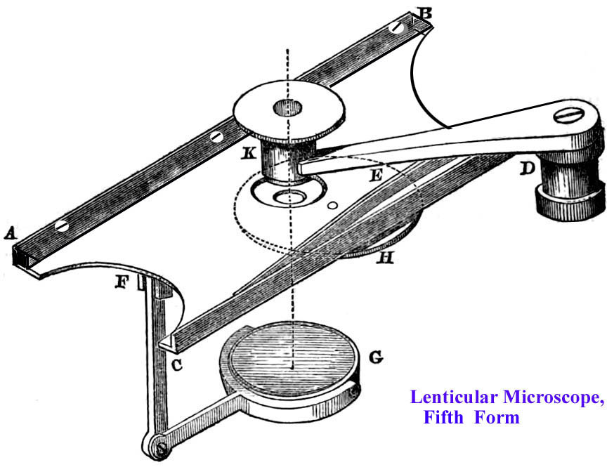
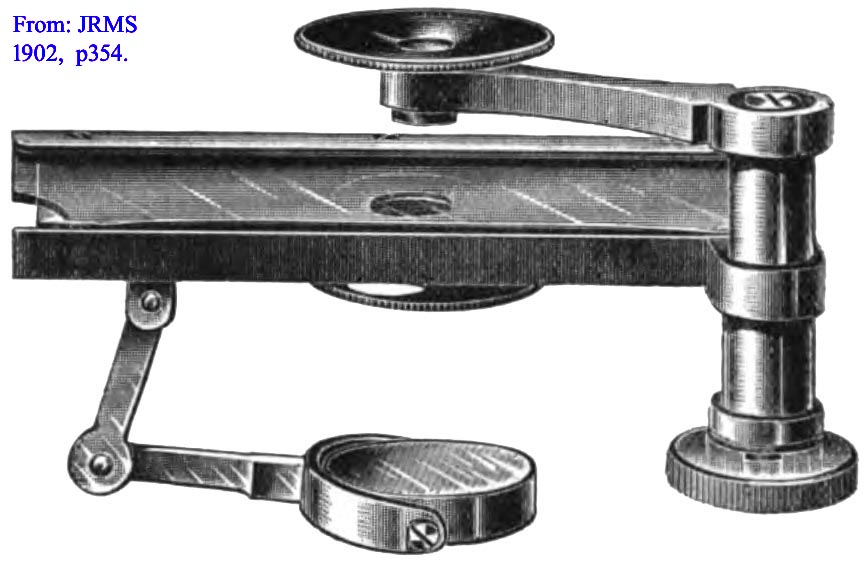 Somewhere between microscopes numbered 53 and 76, the serial number was moved to to the top of the lens arm and both short ends of the stage received curved cutouts. An engraving of this final fifth form, along with a detailed description was reported in the JRMS of 1902, pp353-354.
These microscopes were all said to be supplied with sets of three Coddington lenses. Some sets are known to include 1/4 inch, 1/16 inch, and 1/30 inch focal lengths, and some a 1/4(40X), 1/5(50X), and 1/16(160X). There may have been other magnifications also available. There are four examples of the Pillischer Lenticular in the collection of the Science Museum, London.2 They are all stated to have Wollaston doublet magnifiers, however my research would indicate they are actually Coddington Lenses. I am not certain of how the late Dr Bracegirdle decided that these were all Wollaston doublets rather than Coddington lenses, but he may have assumed they were Wollaston types because their housings are similar to those of actual Wollaston doublets. If the lenses accompanying the Lenticular microscope were Wollaston doublets I would expect the lens side facing the specimen (the object side) to be flat, however the object sides of my examples and that of another example we have examined are all curved as expected for Coddington lenses.
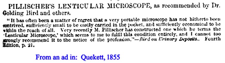 The microscope was advertised in The Lancet of 1853, and the British Medical Directory of 1854. It was described in the entry for 'Microscope' in the 1857 edition of the Encyclopedia Britannica. As noted and pictured above, the Third Form was also illustrated in Golding Bird's fourth edition of Urinary Deposits of 1853. It was also advertised in the back of Quekett's 1855 book3. An excerpt in its entirety from the 1863 edition of Bird is available on this site.
Thanks to Dr Joseph Zeligs, a review of Pillischer catalogs from 1865 to 1872 did not reveal any mention of the Lenticular microscope. Dr Zeligs has also kindly conducted an informal survey of public collections and sales over the years, and along with Paul Ferraglio's review, they note less than fifteen known examples with serial numbers ranging from number 9 to number 439. As I note above, all the known examples with serial numbers above 74, and likely even earlier, are known to be the fifth or final form. A question that may arise is are these serial numbers part of Pillischer's total run of serial numbers or were the Lenticulars numbered separately? A clue has been found by Paul Ferraglio. He found a compound instrument with the same serial number as a lenticular, namely, number 120. This suggests that Pillischer may have actually numbered the Lenticulars separately. Considering the way they are lavishly signed, it would seem that Pillischer was proud of these little microscopes and that might account for the use of a separate run of serial numbers. As shown above, Pillischer apparently ceased production of the Lenticular no later than the early 1860s.
HISTORY OF MICROSCOPES FOR URINALYSIS
Urinalysis is the oldest laboratory test known1. In fact, the Jerusalem proclamation of 1090 stated that any physician who made a diagnosis of a disease without looking at the patient's urine, would be publicly beaten1! Urine microscopy however is a much more recent development, which of course had to wait until the invention of the microscope. Although early microscopists like Leeuwenhoek, reported a few cursory observations of urine under the microscope, serious work did not occur until much later. A great deal has been written about the history of the examination of the urine, quite a bit about the development of urine microscopy2, but almost nothing about the microscopes used or recommended for urinalysis in previous centuries.
Between 1630 and 1783 several microscope users in Europe reported seeing crystals either in voided urine, or in urine that had been allowed to stand at room temperature for a while. It was not until the 1830's when good microscopes were just coming into use, that physicians systematically studied the microscopical appearance of urine. Two Frenchmen were the first to routinely examine their patient's urine under the microscope, and report their findings. These were Pierre Rayer (1793-1867) and his pupil and coworker, Eugene Napolean Vigla, who worked at the Paris Charity Hospital. Their work was reported both in the book 'Traité des Maladies des Reins3' and in journal articles. It included clinico-pathological correlations but did not describe the exact microscope that they used, other than to recommend a microscope of 350X magnification. Rayer's 'Traité des Maladies des Reins4' included illustrations of the important discoveries of cystine crystals (plate 2, figure 6), and red and white blood cells (plate 5) in urine.
In 1841 Alfred Becquerel(1814-1866) published another important book on urine4 in which he, likely for the first time, described dysmorphic red blood cells from patients with the condition we now call glomerulonephritis. Dysmorphic(deformed) erythrocytes are a key marker in the diagnosis of certain specific forms of kidney disease, although their absence is not as specific. The importance of these findings has only recently been rediscovered.
Golding Bird, who published a famous book on urinalysis8-11, was among the first to make recommendations as to the type of microscope that might be useful for this purpose. In his first edition of Urinary Deposits8,' published in 1844, Bird simply recommended a 'vertical instrument on a firm tripod stand, a large ring stage, and a good half inch achromatic' objective. He did point out that a 1/8 inch might be needed for certain fine structures. On the other hand, he noted an experienced observer might only need a half inch objective. In this first edition, Bird did not specify any particular brand of microscope, but did point out that Pritchard of London had constructed a 'microscope of this kind.'
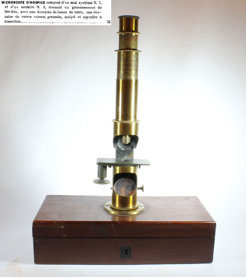 In his second edition(1846)7, Bird noted the relatively inexpensive £7 microscope of Powell and the £5 microscope of Andrew Pritchard, but was more impressed with the advantages and portability of Oberhauser's 'Microscope pour l'Hospice,' a drum microscope.
The recommendation of Oberhauser's model was reiterated in his third and later editions. The manufacture of the 'Microscope pour l'Hospice,' was continued by Oberhauser's successor, Hartnack. An example of this microscope by Hartnack is shown in the accompanying illustration.
In his second edition(1846)7, Bird noted the relatively inexpensive £7 microscope of Powell and the £5 microscope of Andrew Pritchard, but was more impressed with the advantages and portability of Oberhauser's 'Microscope pour l'Hospice,' a drum microscope.
The recommendation of Oberhauser's model was reiterated in his third and later editions. The manufacture of the 'Microscope pour l'Hospice,' was continued by Oberhauser's successor, Hartnack. An example of this microscope by Hartnack is shown in the accompanying illustration.
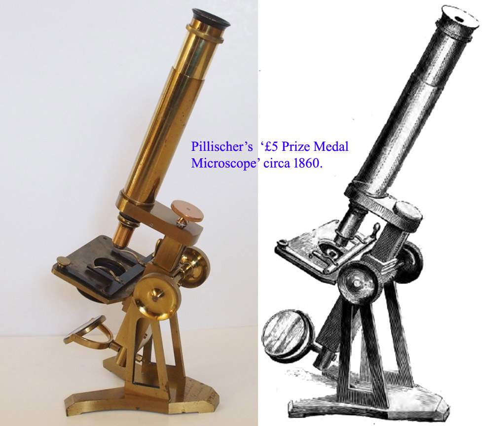 In the third edition11 of Urinary deposits,11 Bird mentions the £7 microscopes of Powell, Smith, and Ross, as well as a £10 one by by Benjamin Dancer. He also states that a good 1/2 inch' objective and 'still better' a 1/4 inch achromatic objective is required. It is in this 18518 edition that he first mentions Moritz Pillischer's compound microscope 'with an excellent quarter inch objective and all other really essential pieces of apparatus' for only £5. Pillischer commenced microscope manufacture about 1848. According to Quekett in his 1852 treatise, Pillischer ranked next in quality after Smith, Ross and Powell & Lealand. Unfortunately, we have not been able to locate any dated catalog or advertisement prior to 1865 which includes a compound Pillischer microscope for £5. As noted above, the Pillischer Lenticular was advertised in the back of the 1855 edition of Quekett's Treatise on the Microscope3, but without a price. In his 1873 catalog, Pillischer offered two microscopes fitting this description, the 'Prize Medal Microscope' and the 'St. Thomas Hospital Microscope.' Both of these came with a quarter inch objective and either one could be purchased for the price which Bird quoted, namely £5. The Prize Medal was awarded during the 1862 London International Exhibition. The St. Thomas Model was not reported until 1871, and so was unlikely to have been available at the time of Bird's original recommendation. One may surmise that it was the Prize Medal Model which Bird was recommending, but it could just as easily have been an earlier undocumented model, though we could find no evidence of an earlier model of Pillischer's priced at only £5.
In 1853, in his fourth edition12, on pages 21-23, after reiterating his comments about Pillischer's compound microscope for £5, Bird recommends, and pictures, for the first time in his books, Pillischer's Lenticular microscope. The image is that of the second form of the instrument, as described above. He spent considerable space extolling the virtues of this little microscope for the examination of urinary deposits at the patient's bedside.
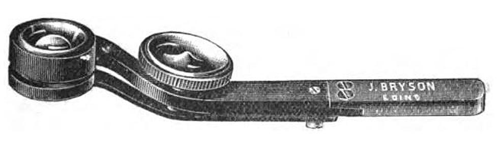
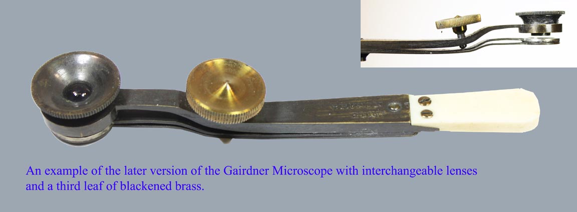 During the same decade, another simple microscope was recommended for urinalysis by Carpenter in his Microscope and its Revelations of 1856. It was invented by Professor William T. Gairdner of Edinburgh and manufactured by J.M. Bryson of the same city. This microscope, as illustrated to the left, did not accept standard slides, had no diaphragms or mirror, and as originally designed, had only one magnification. The lens was a Wollaston doublet. As stated in the JRMS of 1902, Pillischer's model, because of its many superior features, was much more suitable for the purpose of urinalysis. Just like the Lenticular, the Gairdner instrument evolved over the years. The initial instrument, an example of which is in the Golub collection at the University of California, Berkeley, had a fixed single magnifier, but several different instruments were apparently offered, each with its own particular magnification. This original instrument was constructed of two strips of brass, one holding the lens, the other holding a round disk of thin glass. A drop of the liquid specimen would be placed on a separate glass coverslip, which was then placed on top of the fixed glass disk of the microscope with the liquid sandwiched between them. The 'capillary attraction,' (which we now call surface tension), would hold the coverslip in place and allow the microscope to be held up to the light source for observation. The focusing mechanism of this microscope consists of a simple screw on a knob that spreads the two strips of brass apart. Two examples of this original form of Gairdner microscope are in the collection of the Science Museum and are numbers 3/32 and 3/39 in Bracegirdle's CD of that collection. It was suggested that the instrument would be more versatile with removable magnifiers of different powers, and as can be seen in this later example, shown to the right, and kindly provided by Dr Brian Stevenson, this was eventually the way it was supplied. This instrument also possesses another improvement, which is a third brass arm with the coverglass cemented to it with wax.
In reality, as shown by personal observation, these simple microscopes, including the Pillischer Lenticular Microscope show large crystals with ease, but the details of small objects like dysmophic red cells, or cells within casts, would be hard to recognize. It should be noted however, that in Bird's time, crystals remained the most notable and seemingly important findings found under the microscope. In fact, we now know that the majority of crystals which Bird identified are of no pathological significance. Indeed he is often incorrectly credited with the discovery of oxalosis through the observation of calcium oxalate crystals in the urine, or oxalate nephrolithisis for the same reason. This is simply not true13,14,15,16. It is important to note that although Bird observed both red and white blood cells in urine, he failed to make the connection between dysmorphic red cells and Bright's disease (glomerulonephritis). Bird also failed to recognize the frequency and utility of casts in the urine. The identification of the different types of epithelial cells, and the full classification of casts had to wait until much later in the nineteenth century5. Although at the beginning of the twentieth century, most abnormal microscopical findings were identified in the urine17, a full understanding of the significance of most of these did not take place until well into the twentieth century18.
The author is greatly indebted to Dr Joseph Zeligs, Paul Ferraglio, Dr Jurriaan de Groot, Dr Brian Stevenson, Timo Mappes, and James Solliday for their kind assistance, suggestions and contributions to this web page. This page would not be what it is without their help.
REFERENCES AND NOTES
- Offered by Tesseract in catalog 66, in the year 2000
- Bracegirdle, Brian. 'Catalog of the Microscopy Collections at the Science Museum.' Numbers 3/41, 3/44, 3/45, and 3/46. CD-ROM. Little Imp Publications. 2005.
- Quekett, John. Practical Treatise on the Use of the Microscope. 3rd ed., H. Bailliere, 1855.
- Berger, D. 'A brief history of medical diagnosis and the birth of the clinical laboratory. Part 1. Ancient times through the 19th century.' Med Lab Obs, vol.31: 1999, pp. 28-30, 32, 34-40, academia.dk/Blog/wp-content/uploads/KlinLab-Hist/LabHistory1.pdf.
- Fogazzi, G B, et. al. 'The History of Urinary Microscopy to the End of the 19th Century.' Am. J. Nephrol, vol. 14, 1994, pp. 452-457.
- Rayer, P. Traité des Maladies des Reins... Chez J.-B. Baillieà, 1839.
- Becauerel, A. 'Séméiotique des urines, ou traité des altérations de l'urine dans les maladies; suivi d'un traité de la maladie de Bright aux divers áges de la vie.' Fortin-Masson, 1841, p. 505.
- Bird, Golding. Urinary Deposits, their Diagnosis, Pathology, and Therapeutical Indications. 1st ed., John Churchill, 1844.
- Bird, Golding. Urinary Deposits, their Diagnosis, Pathology, and Therapeutical Indications. American ed., Lea & Blanchard, 1845.
- Bird, Golding. Urinary Deposits, their Diagnosis, Pathology, and Therapeutical Indications. 2nd ed.,ition, John Churchill, 1846.
- Bird, Golding. Urinary Deposits, their Diagnosis, Pathology, and Therapeutical Indications. 3rd ed., John Churchill, 1851.
- Bird, Golding. Urinary Deposits, their Diagnosis, Pathology, and Therapeutical Indications. 4th ed., John Churchill, 1853.
- Coe Frederic L., et. al. 'The pathogenesis and treatment of kidney stones.' New Engl. J. Med. vol. 327, no. 16, 1992, pp. 1141-1152.
- Levy F.L., et. al. 'Ambulatory evaluation of nephrolithiasis: an update of a 1980 protocol.' Am. J. Med. vol.98, no. 1, 1995, pp. 50-59.
- Curhan G.C. 'Epidemiology of stone disease.' Urol Clin North Amer.' vol. 34, no.3, 2007, p287-.
- In reality13,14,15, we now know that these crystals are also found in normal urine and that stone formation cannot be predicted either by their presence or absence. Furthermore, calcium oxalate stones are only rarely due to hyperoxaluria(excessive urinary oxalate), but much more commonly due to hypercalciuria(excessive urinary calcium), and sometimes due to hypocitraturia(deficiency of urinary citrate). Primary hyperoxaluria is a very rare condition, particularly in adults. Furthermore, by the time someone develops systemic oxalosis(a disease affecting many different organs), renal failure is often so severe that there are no crystals in the urine at all. Similarly, uric acid crystals do not correlate with uric acid stones. Like some of his contemporaries Bird also erred by confusing the crystals of Calcium oxalate dihydrate, with those of Sodium Chloride (salt). Today we know the only natural crystals diagnostic of specific conditions are cystine crystals (found in cystinuria, a very rare disease), and leucine crystals, sometimes seen in the urine of some patients with liver failure. Today nephrologists also make use of the fact that certain medications and poisons are also associated with specific crystals. Ironically, an unusual type of calcium oxalate monohydrate crystals, in the form of elongated hexagons, which closely resemble hippurate, is found in the urine of patients with ethylene glycol poisoning and this finding is very specific for this type of poisoning.
- Rieder, Herman. Atlas of Urinary Sediments; with Sprecial Reference to their Clinical Significance. as translated by Frederick C Moore and Edited and Annotated by A Sheridan Delepine, Charles Griffin and Co. Ltd, 1899.
- Sobel, Barry. Urinalysis: A Clinicial Guide and Atlas. Published by the Author. 2006.