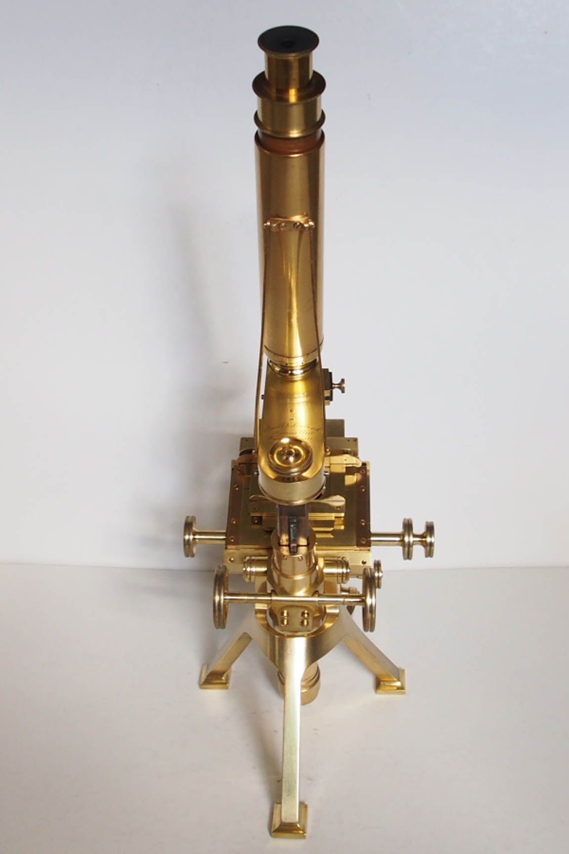
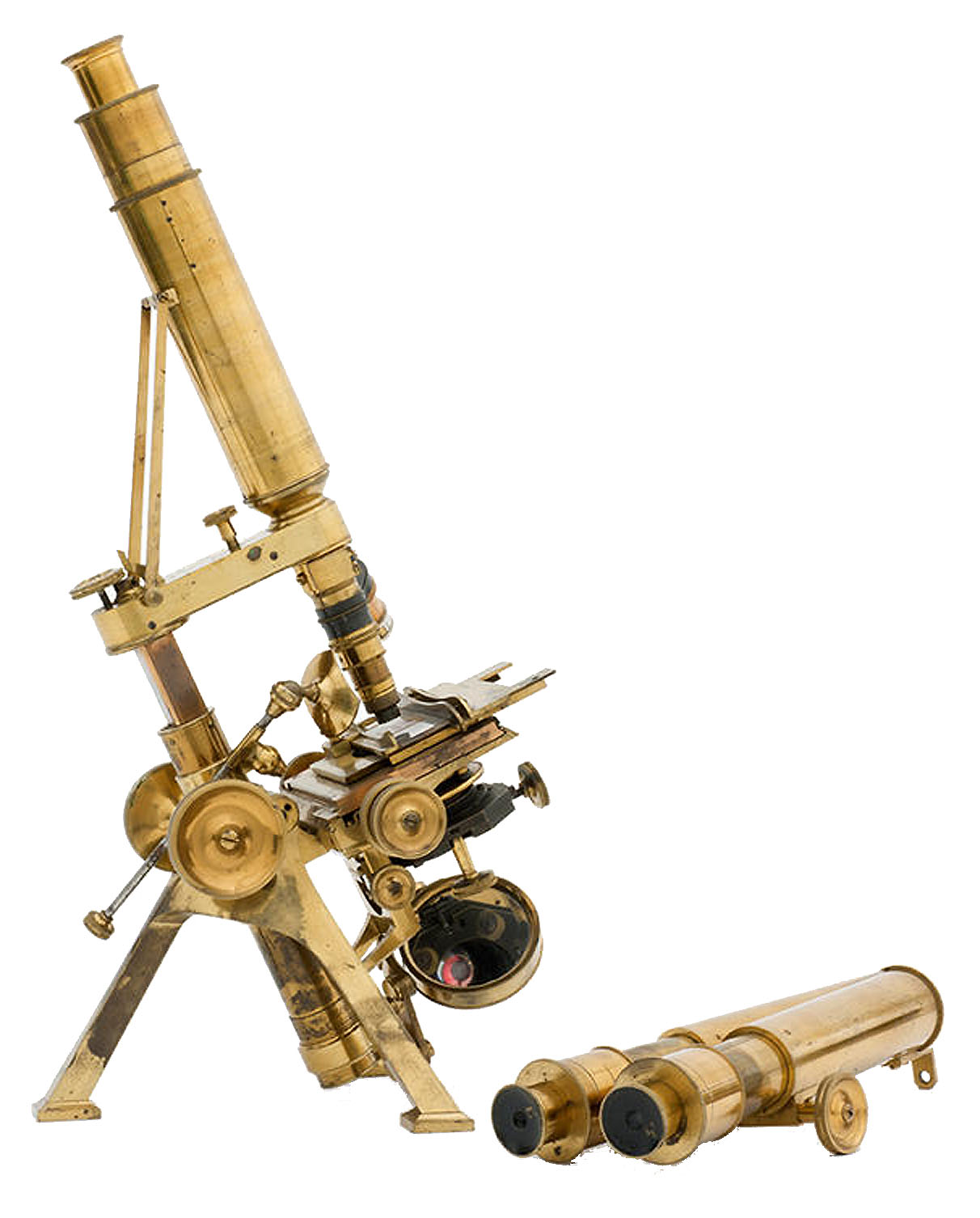
LARGE FIRST CLASS MICROSCOPE
Powell & Lealand, Seymour Place, Euston Square, London, and is dated 1854
| INTRODUCTION | DESCRIPTION | ACCESSORIES | HISTORY |
Please Click On Any Picture for a Larger Version
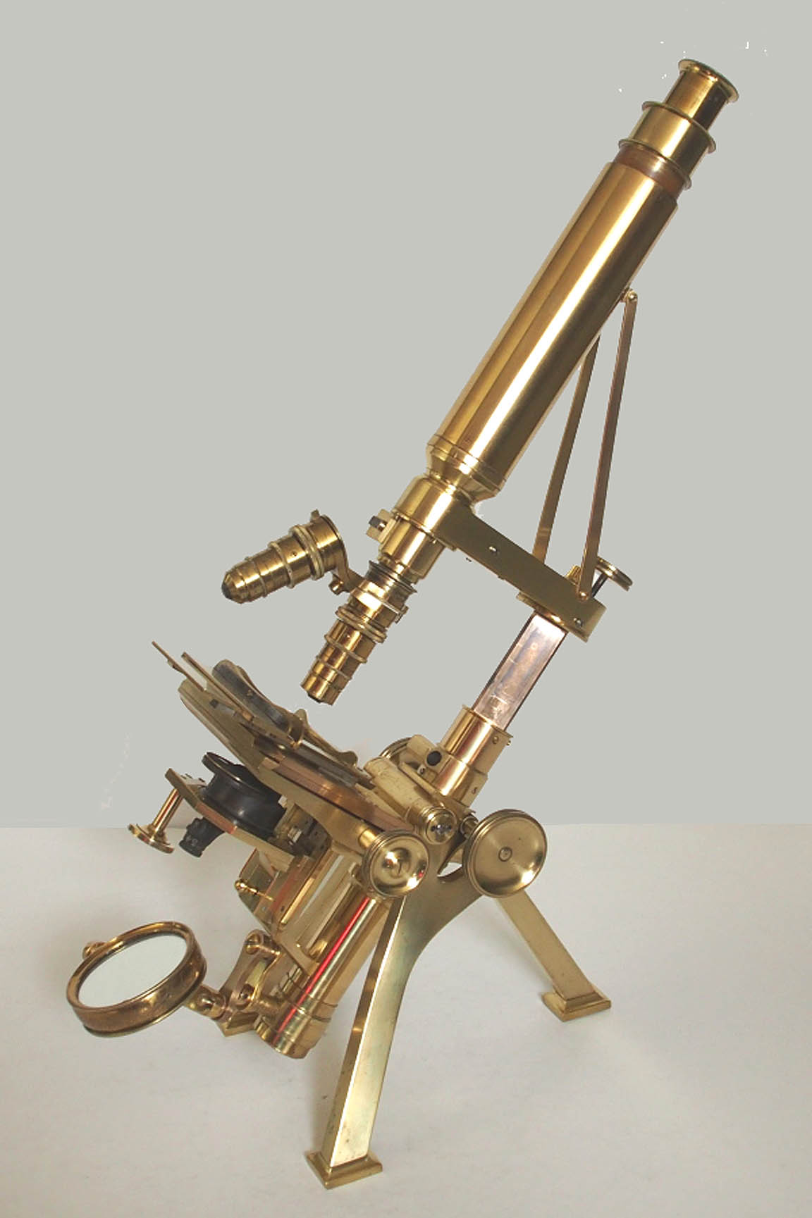
Large First-Class Microscope, which pre-dates the numbering system of models that P & L later used. Another example of this model, dated 1856, is also featured elsewhere on this site. This model represents the small firm's first attempt to develop a larger more solid version of microscope stand based on their
New Microscopeof 1843, and must be regarded as the forerunner of their later No 1 and No 2 stands.
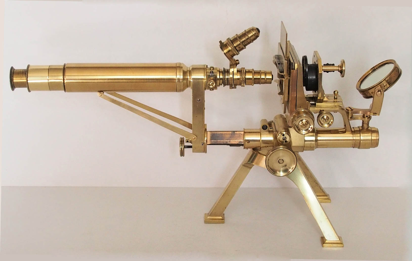
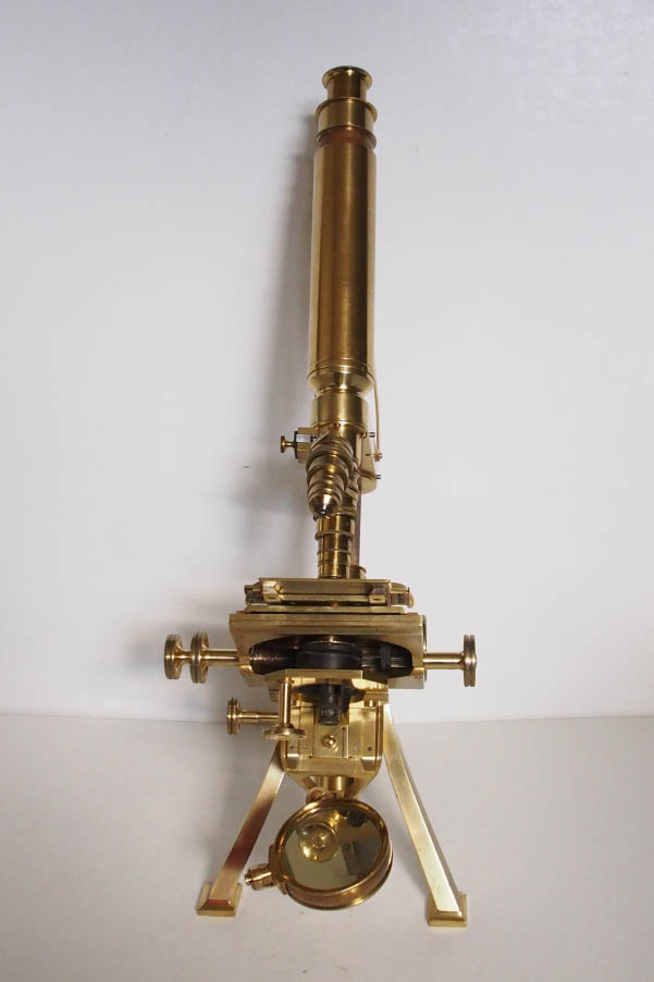
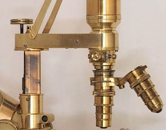 Attached to the bar is the hollow brass arm which houses the fine adjustment mechanism, which consists of a micrometer screw acting on a long lever, which moves the nose-piece. Unlike in another example of this microscope dated 1858, the spring action is here provided by a brass leaf spring acting on the nose-piece instead of the helical coil spring which came into use later. The arm can be rotated sideways through more than 90 deg, with one stop aligning the body tube with the optical axis. It can be fixed into position by a screw which can be tightened with a Tommy-bar.
 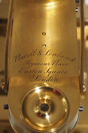 The microscope is signed on the arm:
 Powell & Lealand, Seymour Place, Euston Square, London,and also with the date of
1854. The nose-piece still carries Powell & Lealand's pre-RMS thread, but also present is the firm's own version of the Wenham prism, which slides into the nose-piece, allowing for use of a low-power binocular tube. This modification was introduced in 1861, some seven years after this stand was originally made, and is therefore likely to have been retro-fitted. When purchased, this binocular tube was no longer present.
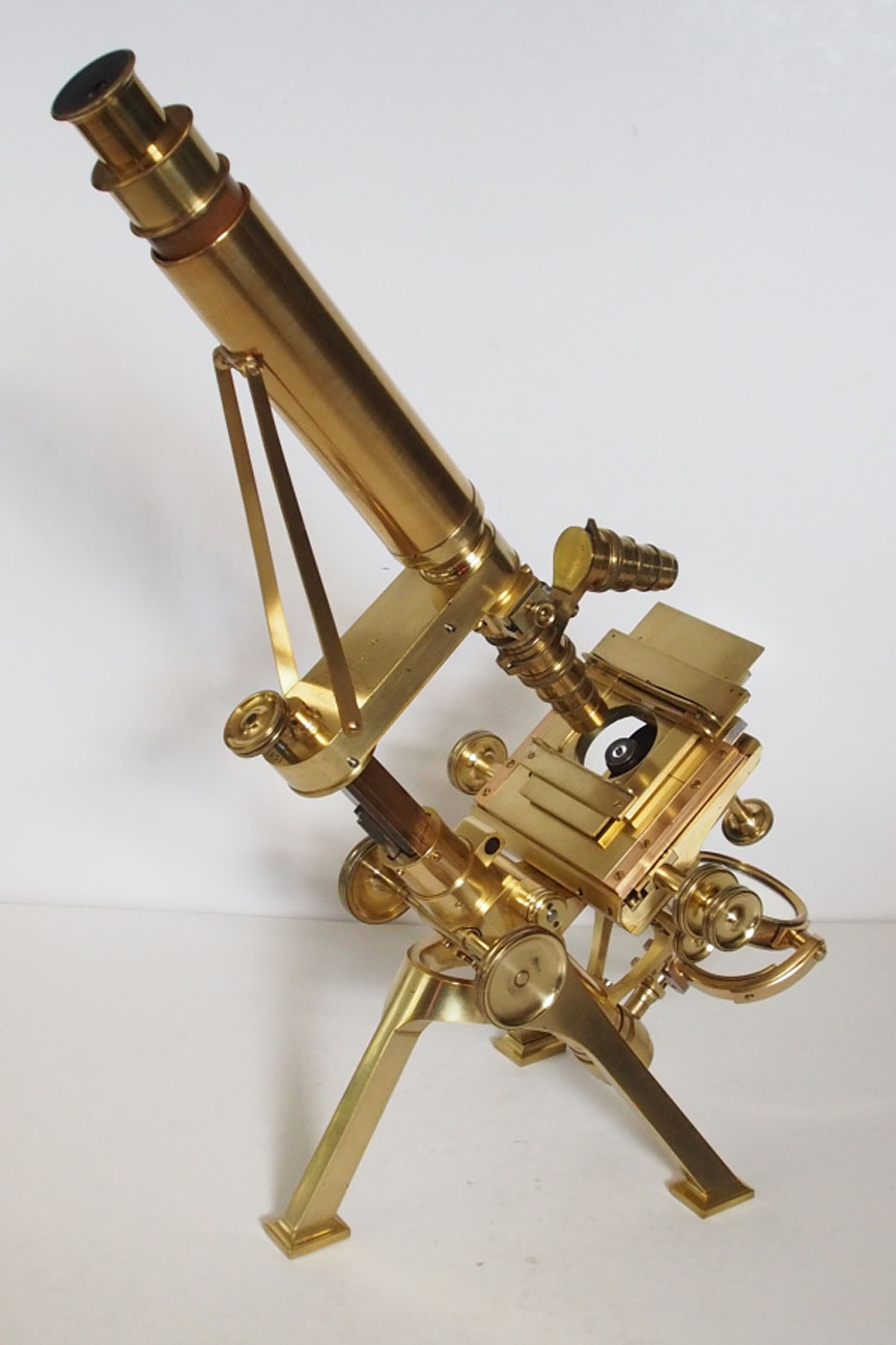
 The Turrell stage (left) is operated through concentric knurled knobs, allowing for a movement of 3/4 inch across the X, and almost 1 inch across the Y axis. The stage plate can be rotated by hand and features a sliding bar with sprung slide supports.
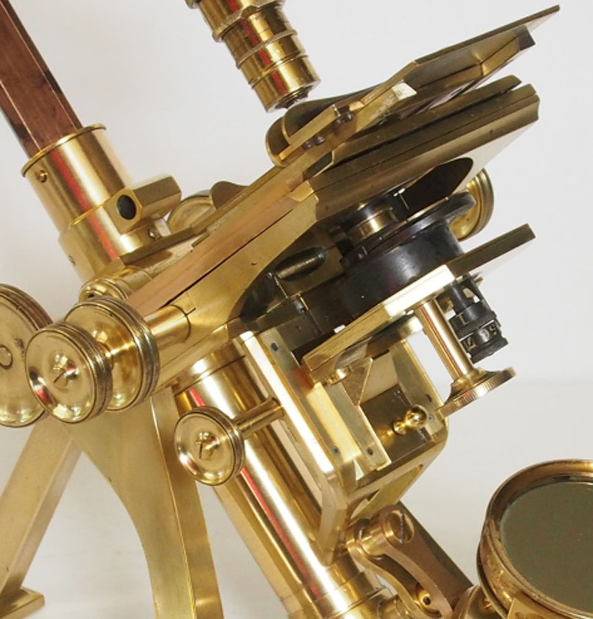 This was the first Powell & Lealand model to feature a separate substage (left) which is moveable by rack and pinion, a departure from the 1843 model, which featured a thicker stage. Attached to the underside of the stage was a fixed brass plate with bayonet mount for substage accessories. On these earlier models without a separate substage, the condenser was fitted with its own intrinsic focusing mechanism.
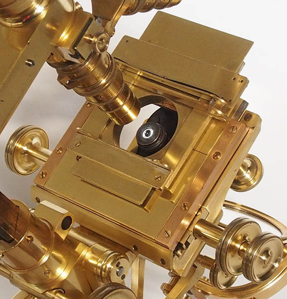
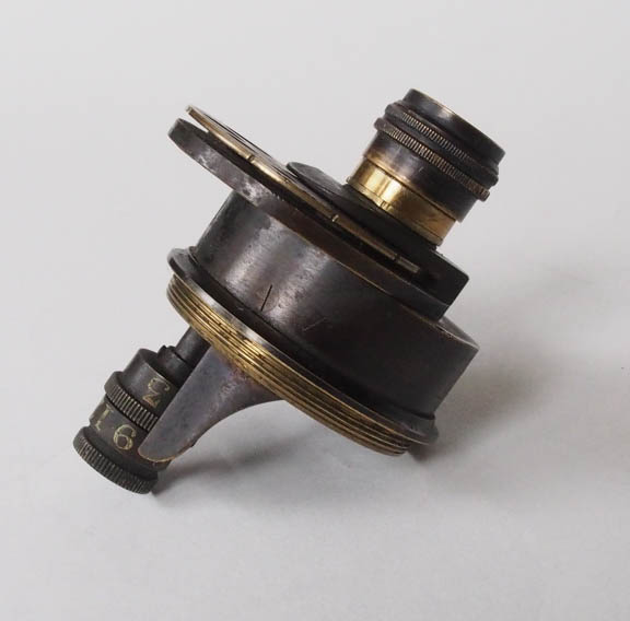
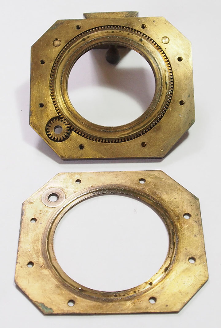 On the 1854 model featured on this web page, substage accessories are screwed into a threaded ring, which can be rotated by a vertically placed knurled knob, which activates a pinion acting on a ring gear, which are both hidden inside the substage plate(shown disassembled to show the mechanism). Unusually, this example is not as yet fitted with the pair of screws set at right angles for centering the substage along its X and Y axes, as seen in examples from 1855 onwards. Instead, this is achieved by loosening the 2 screws which affix the substage plate to the dovetailed slider, thus aligning the condenser stops with the optical axis. This suggests that this particular example was made at an earlier stage of development, and indeed, no examples of this model are known to exist carrying an earlier date. The dovetailed slider allows for easy removal of the substage assembly without affecting the alignment, and can be replaced with another slider carrying, for example, an aperture for insertion of dark wells. In this example, no additional slider is present.
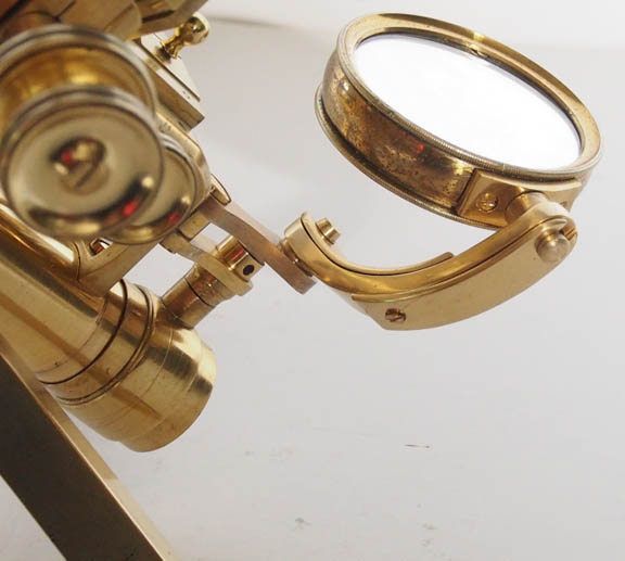 The plano-concave mirror is mounted on a single quadrant gimbal, the attachments of which are sprung. This is carried on a double articulated arm which is attached to a collar running on the tail-piece of the lower limb. The microscope stands some 18.1 inches (460 mm) tall when in use. There is no case.
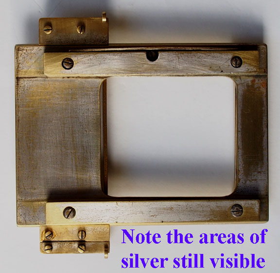 On close inspection, as shown to the right, it appeared, that the top stage plate showed signs of having been silvered. This is most apparent on its under surface, and after cleaning, this was left as is. This finding may be of relevance when considering the origins of this microscope(see, Possible Origins, below).
 Powell & Lealand's New Microscope. The original form is supported by a lighter version of the tripod, which carries the body on trunnions, and a transverse bar, which contains a long lever, which moves the nose piece fine-focus motion. Initially, the fine-focus screw, which operated a cone, was placed on the right-hand side. In 1848 this was moved to the familiar position on the top of the bar, immediately behind its pivot. From the outset, the microscope tube was also supported by a set of diagonal supporting struts extending from the back of the bar to the upper third of the tube. Sometime around 1860 these were abandoned, when an exchangeable binocular tube to the design of Francis Wenham was offered. This rendered the struts an encumbrance, and it was also apparent that they were doing little to improve stability.
The next microscope made by Messrs. Powell & Lealand is not, I believe, figured or described anywhere; neither can we now assign a precise date for its introduction. I have asked Mr. Thomas Powell about it, and he tells me his own instrument is dated 1855, and that, as it was only worked at in odd times, it took a long time to make. It is not improbable therefore that this model was brought out after the Exhibition of 1851. It will be unnecessary to illustrate this instrument, as it is very similar to the No 1 stand as at present made. The differences between them are as follows: - the tripod foot is smaller, but similar in every other respect; the stage has non-concentric rotation; in the substage the pinion for rotary motion is placed in a vertical instead of horizontal position as at present, and the substage apparatus screws into the substage. The back-stay fitted to the bodies of previous models is omitted. This was the first microscope made by this firm that had a complete substage; in this point it was probably a copy of the Microscope Andrew Ross prepared for the exhibition of 1851, at which Messrs. Powell & Lealand were not exhibitors.
For the sake, however, of such as may desire the power of obtaining a more oblique illumination, than is permitted by the construction of the stage in the instrument just described, Messrs. P & L have recently brought out a new pattern, in which the thickness of the stage is greatly reduced, a sub-stage is provided for the reception of the condenser and other fittings, and the mirror is mounted on a double-extending arm.
Powell & Lealand's improved microscopecomplete with Amici prism on stand, clearly identical to the model featured here, and with a full description:
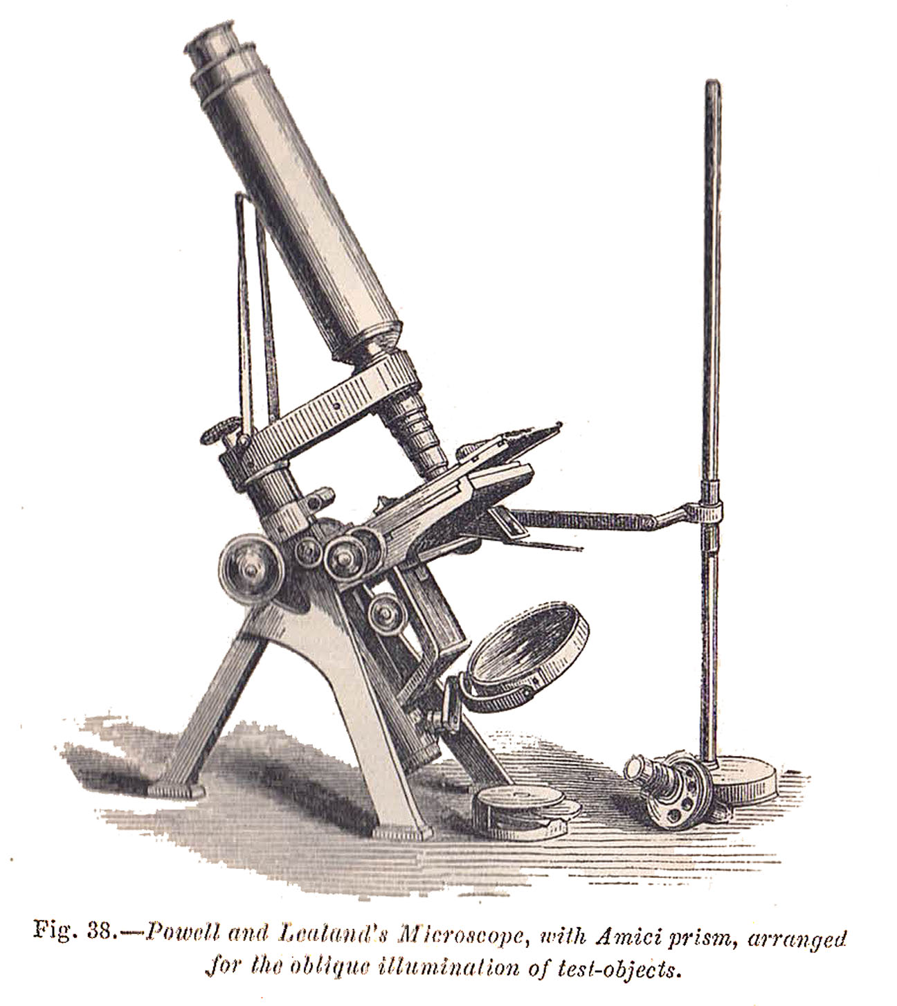
The three legs are considerably stouter and more inclined than in their former instrument, which gives additional stability; they support, at their upper part, the trunnions to which the tube and the stage are attached. From out the tube a triangular bar is raised by a rack and pinion connected with the milled head. To the upper part of the triangular bar a broad arm is fixed, bearing the compound body; this arm is hollow, and contains the mechanism for the fine adjustment, which is effected by turning the small milled head. The arm is connected with the triangular bar by a strong conical pin, on which it turns, so that the compound body may be moved aside from the stage when necessary; by a mechanical arrangement it stops when central. The stage is of an entirely new construction, having vertical, horizontal, and circular movements, and graduated for the purpose of registering objects so as to be found at pleasure; and in order to do this effectually a clamping piece is provided against which the object slide rests, and the circular motion is stopped. It is an exceedingly effectual method of finding any favourite object. The stage is remarkably strong, and at the same time so thin, that the utmost obliquity of illumination is attainable, the under portion being entirey turned out: it has a dove-tailed sliding bar moveable by rack and pinion, on turning the milled head into this bar slides the understage, having vertical and horizontal motions for centering, and also a circular motion; into the stage are affixed the various appliances for underneath illumination. The achromatic condenser, if of 100 deg. of aperture, with nine apertures and five central stops, the apertures and stops having independent movements, the manipulator can regulate at will; this is considered to be a great improvement. There is an appliance provided for the dark-wells, which is put into the dove-tailed sliding bar instead of the underneath stage. The mirror is attached to a quadrant of brass and two arms, in order to obtain greater obliquity of illumination; the whole fits into a short piece of tube made to slide either up or down the long tube attached to the bottom of the stage by which the mirror is connected with the other part of the stand; the reflectors themselves are both plane and concave, as in other instruments. The achromatic prism for oblique light is very useful for bringing out the fine markings of the most difficult Diatomaceae with the high-power object glasses. This instrument combines extreme steadiness with remarkable simplicity, together with every motion and appliance that has ever been discovered for the microscope; the compound body is supplied with a draw tube. Messrs. Powell and Lealand microscopes are sold at prices suitable to the wants and means of most persons; their No 1, such as we have represented, but without object-glass can be purchased for 22 Pounds. A smaller instrument, fit for the student, with 3/4 inch of motion to the stage by means of a lever, coarse and fine adjustments to body, plane and concave mirrors, revolving diaphragm, Lister's dark-wells, and two eye pieces 8 Pounds.Incredibly, the engraving of this model P & L microscope continues to be included in subsequent editions of this popular work on the microscope, right until the last printing of the 15th edition (1911), with the erroneous subscript:
Powell and Lealand's Student's microscope. It would appear, that this model continued to be produced throughout the 1850's in parallel to what was later to become the
No 3 stand, from which it originated. Throughout this period, both types would go through their own stages of development (see Budd J LaRue's articles on the P & L No 2 and No 3 microscopes). Careful examination of extant examples shows both the point at which these two models diverged, and how each subsequently started to resemble the later No 2 and No 3 stands into which they evolved. The example featured on this page thereby seems to be the
missing link, and can be dated to shortly after this point of divergence occurred. (see Table 1, below)
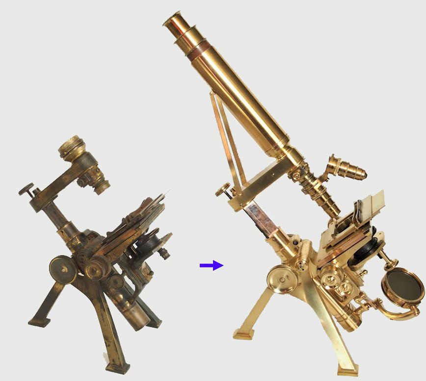
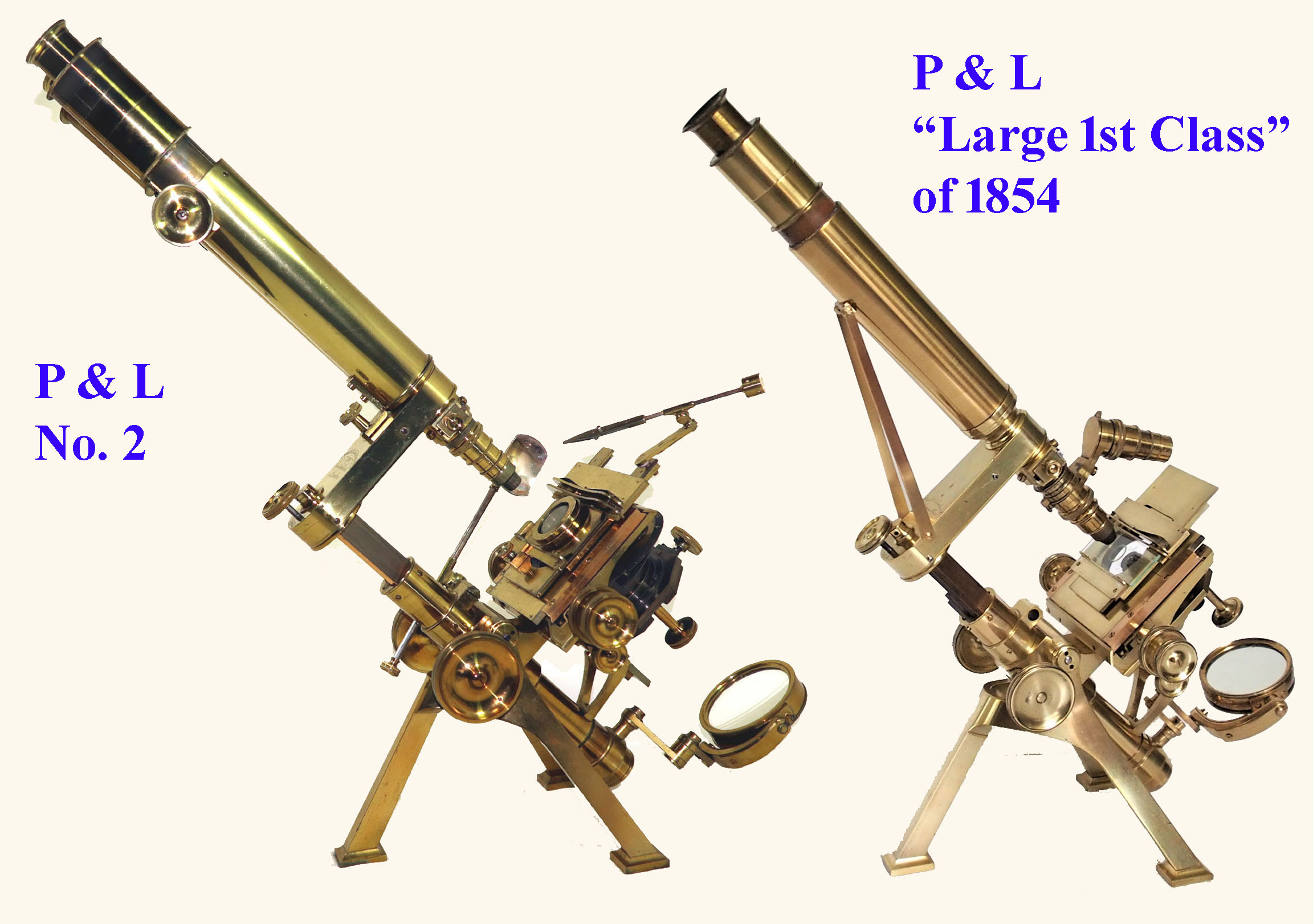 These images allow for a comparison between the Powell & Lealand improved Large First Class Microscope dated 1854, and an example dated 1862 of the later Powell & Lealand No 2 stand into which it evolved. By 1860 the firm had developed a successor to their first class microscope, from then on referred to as their No 1 model, but it was decided to keep producing the original improved model alongside this as a slightly less elaborate option, with a few modifications, thereafter referred to as their No 2 microscope stand.
| DATE | SOURCE | ADDRESS | FEATURES |
|---|---|---|---|
| 1848 | HSM Collection, London | 4 Seymour Place, Euston Square, London | Braces to body tube Side arm fine focus Cylindrical coarse focus housing Thick stage Plug substage Tripod 1 |
| 1849 | Wellcome Collection | 4 Seymour Place, Euston Square, London | Braces to body tube TOP OF ARM FINE FOCUSING Cylindrical coarse focus housing Thick stage Plug substageTripod 1 |
| 1850 | Website of Allan Wissner | 4 Seymour Place, Euston Square, London | Braces to body tube Top of arm fine focus Cylindrical coarse focus housing Thick stage Plug substage Tripod 1 |
| 1851 | HSM Collection | 4 Seymour Place, Euston Square, London | Braces to body tube Top of arm fine focus Cylindrical coarse focus housing Thick stage Plug substage Tripod 1 |
| 1852 | Frank Collection | 4 Seymour Place, Euston Square, London | Braces to body tube Top of arm fine focus Cylindrical coarse focus housing Thick stage Plug substage Tripod 1 |
| 1853 | RMS Collection as part of the HSM | 4 Seymour Place, Euston Square, London | Braces to body tube Top of arm fine focus Cylindrical coarse focus housing Thick stage Plug substage Tripod 1 |
| 1854(a) | Collection of Brian Stevenson | 4 Seymour Place, Euston Square, London | Braces to body tube Top of arm fine focus Cylindrical coarse focus housing Thick stage Plug substage Tripod 1 BUT WITH SMALL RECTANGULAR FOOT PADS |
| 1854(b) | Example featured on this page | 4 Seymour Place, Euston Square, London | Braces to body tube Top of arm fine focus HEXAGONAL COARSE FOCUS HOUSING, CYLINDRICAL AT TOP THIN STAGE SEPARATE SUBSTAGE WITH ROTATION BUT NO CENTERING FOR THE SUBSTAGE Tripod 2 |
| 1855 | Sotheby's Catalog Oct 7, 1994 Lot 325. |
4 Seymour Place, Euston Square, London | Braces to body tube Top of arm fine focus Hexagonal coarse focus housing, cylindrical at top Thin Stage Separate substage with rotation but now WITH X-Y CENTERING FOR THE SUBSTAGE Tripod 2 |
| 1856 | This siteFormer Collection Barry Sobel | 4 Seymour Place, Euston Square, London | Braces to body tube Top of arm fine focus Hexagonal coarse focus housing, cylindrical at top Thin Stage Separate substage with rotation with X-Y Centering for the substage Tripod 2 |
| 1857 | Frank Collection | 170 EUSTON ROAD, London | Braces to body tube Top of arm fine focus Hexagonal coarse focus housing, cylindrical at top Thin Stage Separate substage with rotation with X-Y Centering for the substage Tripod 2 |
| 1858 | Golub Collection | 170 Euston Road, London | Braces to body tube Hexagonal coarse focus housing, cylindrical at top Thin Stage Separate substage with rotation with X-Y Centering for the substage Tripod 2 |
| 1859 | Sotheby's Catalog Oct 7,1994 Lot 375 |
170 Euston Road, London | Braces to body tube Hexagonal coarse focus housing, cylindrical at top Thin Stage Separate substage with rotation with X-Y Centering for the substage Tripod 2 |
| 1860 | Sotheby's Catalog Oct 7,1994 Lot 326 |
170 Euston Road, London | FIRST EXAMPLE OF NEW No 1 MODEL: NO BRACES Top of arm fine focus Thin stage WITH CONCENTRIC ROTATION WITH STAGE ABOVE STAGE-SUPPORT RING RECTANGULAR PRISM-SHAPED COARSE FOCUS HOUSING Complete Separate substage with rotation with X-Y Centering for the substage LARGER VERSION OF Tripod 2 |
| 1861 | Golub Collection | 170 Euston Road, London | No 2 MODEL: No braces Top of arm fine focus HEXAGONAL coarse focus housing, CYLINDRICAL AT TOP Thin stage with concentric rotation with stage above stage support ring. Complete Separate substage with rotation with X-Y Centering for the substage Larger version of tripod 2 |
| after 1862 | Article: The P & L No 2 Microscope (see bibliography below) |
170 Euston Road, London | 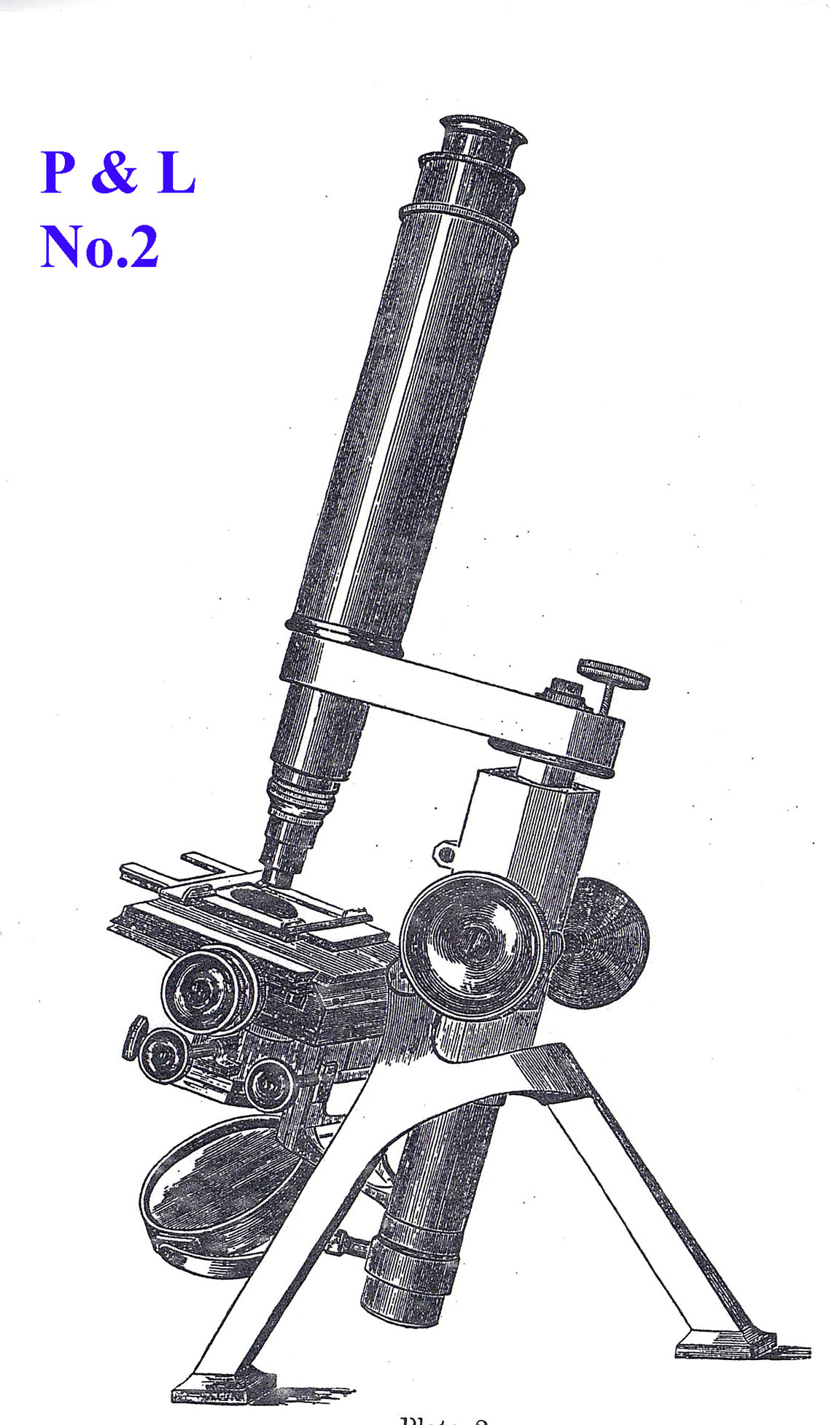 No 2 MODEL: No 2 MODEL:No braces Top of arm fine focus HEXAGONAL COARSE FOCUS HOUSING ALL THE WAY TO THE TOP Thin stage with concentric rotation with stage above stage support ring. Complete Separate substage with rotation with X-Y Centering for the substage, BUT CONTROL KNOB FOR ROTATION NOW ORIENTED HORIZONTALLY. Larger version of tripod 2 |
 Tripod 1: Rank feet with small circular foot pads as in later No 3 stand
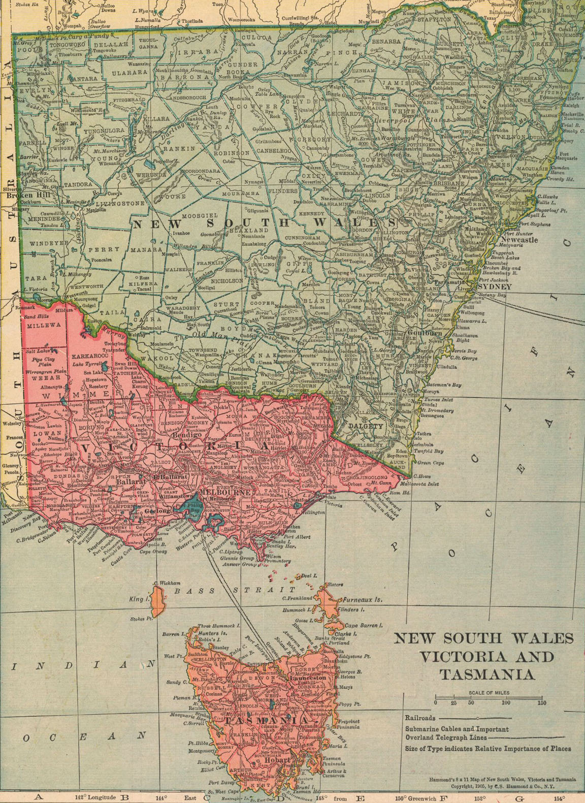 This microscope was purchased on eBay from a vendor in Tasmania, Australia, who had bought it from someone who had acquired it at an auction, either in Tasmania, or possibly on mainland Victoria. Otherwise, its true origins are unknown, but it is an interesting exercise to give in to some speculation at this point, and take a short historical excursion. Microscopy, and indeed science, in these distant outreaches of the British Empire remained the precinct of amateurs for most of the 19th century, many of them belonging to other professional groups, such as medicine, pharmacy, but also the clergy. At first, informal networks formed of like-minded enthusiasts, but soon, to develop into Societies, including Microscopical- Naturalist-, or local Royal Societies.
Reflecting on Tasmania, and then on microscopy, the name of Dr. William Valentine (1808-1876) comes to mind. Originally from Nottingham, he commissioned
Andrew Ross in 1831 to construct one of his earliest achromatic microscopes. Dr. Valentine emigrated to Tasmania in 1840, becoming assistant surgeon to the Campbell town district. On such a small island, word of Valentine's scientific knowledge and his magnificent microscope (described as the finest in the colony)
spread quickly, and his house - The Grange, in Campbelltown, which he commissioned in 1847 - became a hub for naturalists. There is no evidence, however, to show that Valentine ever owned a Powell and Lealand microscope. In 1877, so shortly after his death, the Proceedings of the Royal Society of Tasmania report:
This microscope was purchased on eBay from a vendor in Tasmania, Australia, who had bought it from someone who had acquired it at an auction, either in Tasmania, or possibly on mainland Victoria. Otherwise, its true origins are unknown, but it is an interesting exercise to give in to some speculation at this point, and take a short historical excursion. Microscopy, and indeed science, in these distant outreaches of the British Empire remained the precinct of amateurs for most of the 19th century, many of them belonging to other professional groups, such as medicine, pharmacy, but also the clergy. At first, informal networks formed of like-minded enthusiasts, but soon, to develop into Societies, including Microscopical- Naturalist-, or local Royal Societies.
Reflecting on Tasmania, and then on microscopy, the name of Dr. William Valentine (1808-1876) comes to mind. Originally from Nottingham, he commissioned
Andrew Ross in 1831 to construct one of his earliest achromatic microscopes. Dr. Valentine emigrated to Tasmania in 1840, becoming assistant surgeon to the Campbell town district. On such a small island, word of Valentine's scientific knowledge and his magnificent microscope (described as the finest in the colony)
spread quickly, and his house - The Grange, in Campbelltown, which he commissioned in 1847 - became a hub for naturalists. There is no evidence, however, to show that Valentine ever owned a Powell and Lealand microscope. In 1877, so shortly after his death, the Proceedings of the Royal Society of Tasmania report: The Secretary informed the meeting, that the microscope then on the table was presented to the Society by Mr. P. T. Smith prior to his departure for England. The instrument was a first class-one by Ross, and was furnished with very many accessories, a vast number of mounted objects, etc.. It was a very great acquisition for the Society, as its value (with numerous appliances) could not be far off GBP 200. It had recently belonged to the late Dr. Valentine of Campbell Town. Dr Valentine must therefore have remained faithful to Ross instruments. It is also unknown what type of microscope was used by his friend and collaborator, Ronald Campbell Gunn (1808-1881), with whom he used the microscope to identify a moss (Splanchnum Sphaericum), that Valentine had collected growing on the bones of an escaped convict in 1845.
 A very similar Powell and Lealand microscope to the one described here is present in the Medical History Museum at the University of Melbourne, once belonging to Professor George Britton Halford (1824-1910). This also features the stabilizing struts, and rectangular section legs, and the museum's inventory reports that:
It is stored in a walnut case with side compartments, and includes inventory and usage notes by its user. In 1864 the University of Melbourne bought the microscope for Dr. G.B. Halford from the Reverend John Bleasdale, a prominent Catholic clergyman and an active member of the Microscopic Society of Australia. Made by Powell and Lealand, London, the stand has been made 'proof against corrosion of acids and the injurous actions of alkalis and spirit of wine by plating the brass with silver and then gilding with more than half an ounce of the purest gold'. The donor was the grand-daughter of G.B. Halford.Pre-dating 1860, this microscope is likely to have been retro-fitted with the firm's low-power binocular prism and Wenham binocular body tubes some years later. The University obviously sought to impress Dr Halford, who obtained his medical degree at St George's Hospital in London, and was to become the first full-time Professor of Medicine, Anatomy, and Physiology at the newly established Medical School. Another, perhaps relevant, piece of the puzzle is provided by an article in the Argus newspaper of Melbourne of Thursday, 22 July 1858:
The Microscopic Society held its usual monthly meeting on Tuesday evening, at the residence of Mr. W. S. Gibbons, the honorary secretary*. There was a good attendance of members,and no fewer than seven microscopes, most of them first-class instruments, occupied the tables. Messrs. W. B. Jackson and W. Potts were admitted members. Mr. Jones submitted a new instrument by Powell and Lealand, with several interesting objects, just arrived. Mr. Bleasdale produced crystallised iodides of some of the alkaloids. Mr. Gibbons showed a collection of fossil sections, illustrative of the coal and other formations; and also a suite of micro-photographic apparatus, which he had lately imported from London. It was arranged that the next monthly meeting should be held on Thursday, the 17th proximo, at the residence of Dr. Eades. The meeting was occupied in the late hour in the observation and discussion of the instruments and objects submitted; and agreed to hold an extra meeting on Friday evening (tomorrow), at the residence of Mr. Bleasdale, for the purpose of receiving a paper by Mr. Ralph on the construction of glass cells.Could the 1854 Powell and Lealand microscope featured on this web page have been Mr. Jones'? It would have been 4 years old in 1858, and therefore not new. Was it Mr. Jones's instrument, which Bleasdale sold in 1864 to the University (and he mediated in the sale), or was it one of his own, given its silver/gold plating, it having been used for his own favourite pastime, the observation of crystals of corrosive chemicals? As mentioned earlier, the example under discussion here, and dated 1854 shows signs of the stage having been silver plated. This, and the rest of the body were in almost black oxydised condition when found. Could this have been the effect of damp conditions in which the microscope - now a hack - was kept by successive owners, or were
iodides of alkalisto blame, and this microscope once belonged to the Rev. John Ignatius Bleasdale(1822-1884)?
In the early days of Melbourne something of a sensation was caused when it was announced that Mr Gibbons was importing a microscope at a cost of more than 100 Pounds for his professional use. It went down in a wrecked ship on the way out, however, and a second instrument, still more valuable, was obtained". Gibbons was a Fellow of the Chemical Society and a Fellow of the Royal Microscopical Society, and owned a most interesting collection of minerals. The same article mentions that his death at the age of 92 was "the result of a cable tram accident which occurred on the evening of July 13, when he was struck by a car while crossing the tramway track.