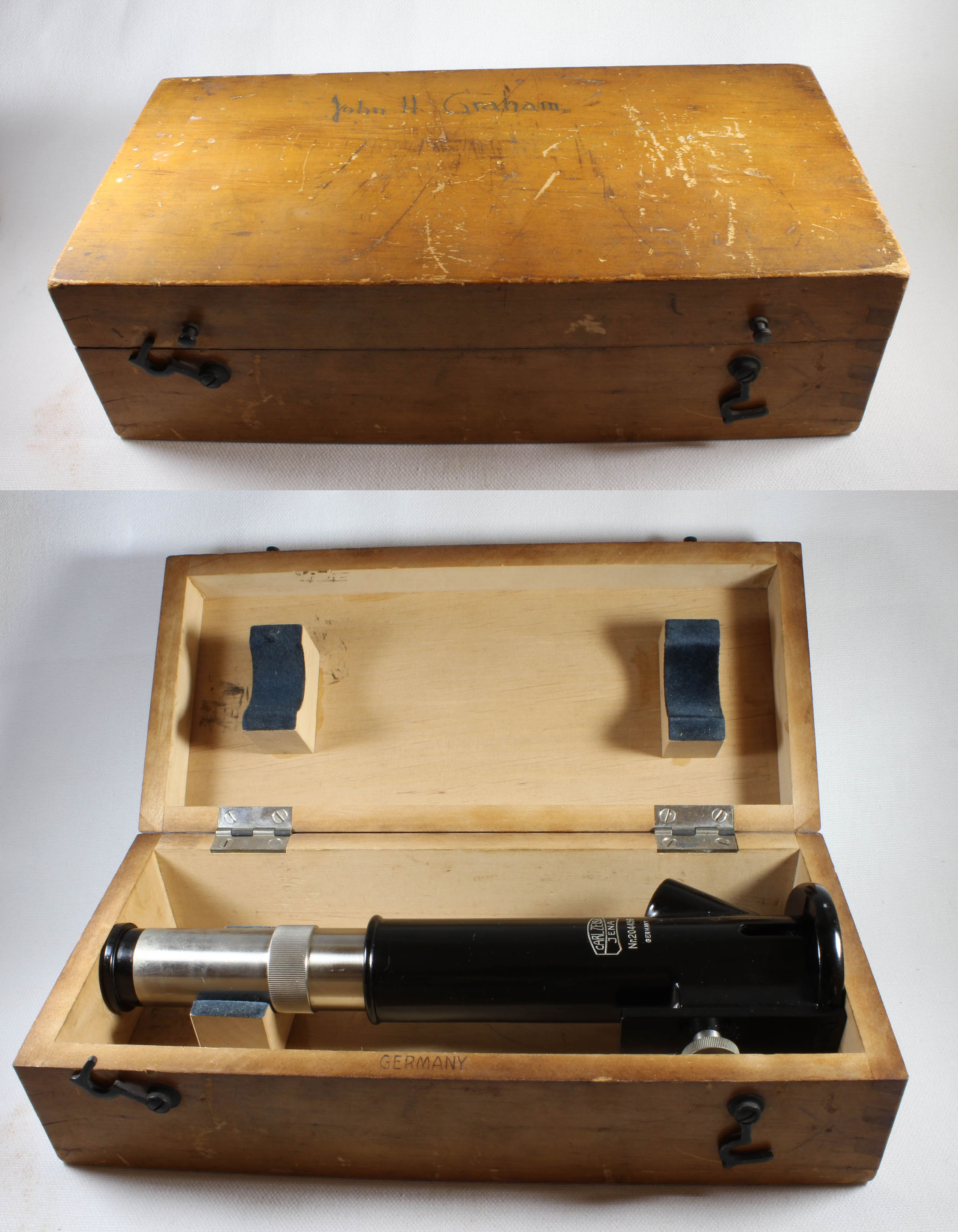
MICROSCOPE-ANTIQUES.COM © 2013-15.
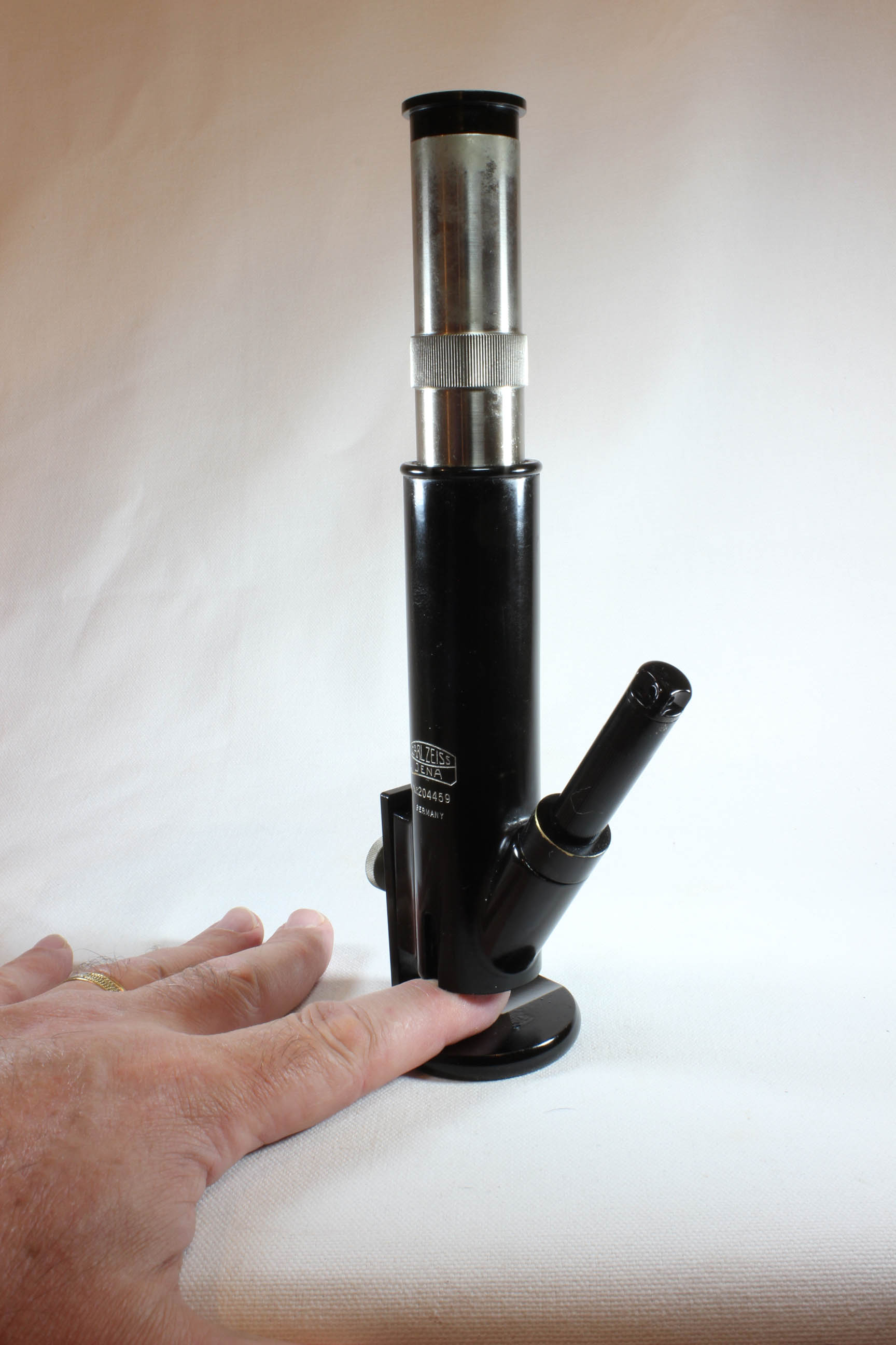
MAKER: CARL ZEISS OPTISCHE WERKSTATAETTE
MODEL: 'SKIN MICROSCOPE' or 'SMALL MULLER SKIN MICROSCOPE'
c. 1928
SIGNED: Carl Zeiss, Jena
SERIAL NUMBER: 204459
Please Click On Any Picture for a Larger Version
DESCRIPTION:
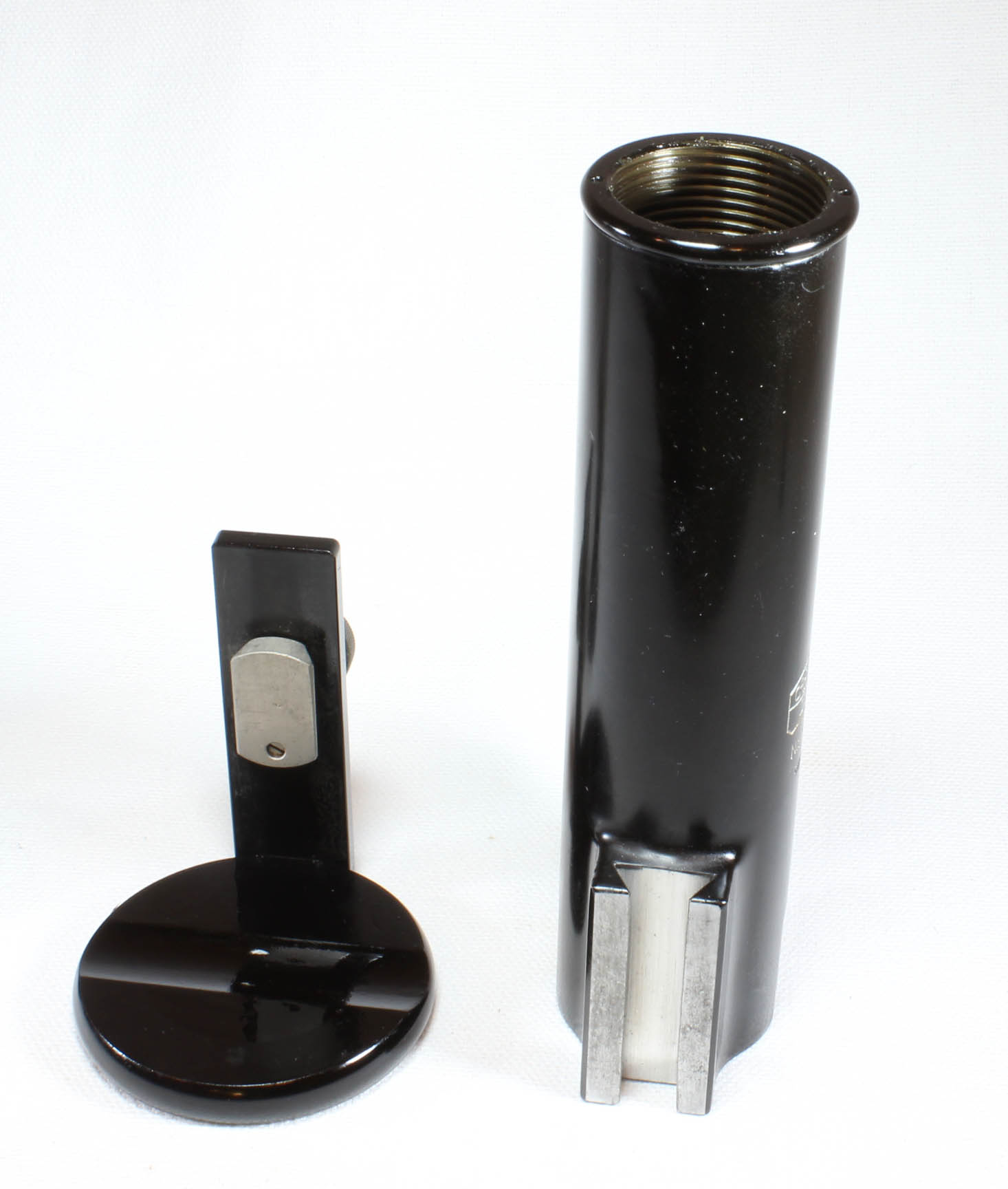
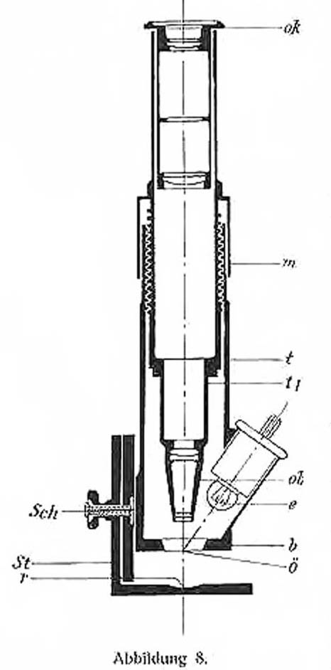
The microscope is a modified drum-type. The foot is round and black and has a semicircular depression for the subject's finger. A metal rail arising from the foot forms the vertical support for the microscope via a sliding dovetail fitting; coarse focus is effected by sliding the instrument up or down on the dovetail, and then using the setscrew on a knob to lock it in place. Fine focus is achieved by rotation of the body tube. The instrument has a 10X eyepiece and a 6X objective giving a final magnification of 60X. A port angled towards the center of view comes in from the side. This is designed to allow a light source to be placed inside it. This kind of angular top-lighting allows good visualization of the skin withut glare or undue reflection. The original light source, (no longer present) would have been powered by a pair of wires attached to a separate battery, or as the instructions say, via a resistor to a higher voltage source such as AC household current. In use, a finger is placed in the depression on the foot, the skin of the base of the nail ('nail fold') is covered with oil (to act as a clarifying agent) and the microscope is gently lowered until just touching the skin. Fine focus then allows visualization of the capillaries, though critical focus requires the subject to hold very still. This instrument has a narrow depth of focus; this is a disadvantage as discussed below.
HISTORY OF THIS MICROSCOPE AND SKIN CAPILLARY MICROSCOPY
 The idea of studying capillaries under the
microscope dates back many centuries. In 1628 William Harvey described the circulation of blood, and the heart
as its pump. Harvey did not visualize the capillaries, instead referring to 'pores.' It was not until 1661 that
Marcello Malpighi first visualized, via a microscope, the movement of red blood cells through capillaries of the
lung and the urinary bladder of a frog. In 1663 Johan Kolhaus was apparently the first to visualize the
capillaries of the nail fold. The use of oil to make the skin over the capillaries in this area more transparent
was apparently first suggested by Unna in 1893.
The idea of studying capillaries under the
microscope dates back many centuries. In 1628 William Harvey described the circulation of blood, and the heart
as its pump. Harvey did not visualize the capillaries, instead referring to 'pores.' It was not until 1661 that
Marcello Malpighi first visualized, via a microscope, the movement of red blood cells through capillaries of the
lung and the urinary bladder of a frog. In 1663 Johan Kolhaus was apparently the first to visualize the
capillaries of the nail fold. The use of oil to make the skin over the capillaries in this area more transparent
was apparently first suggested by Unna in 1893.
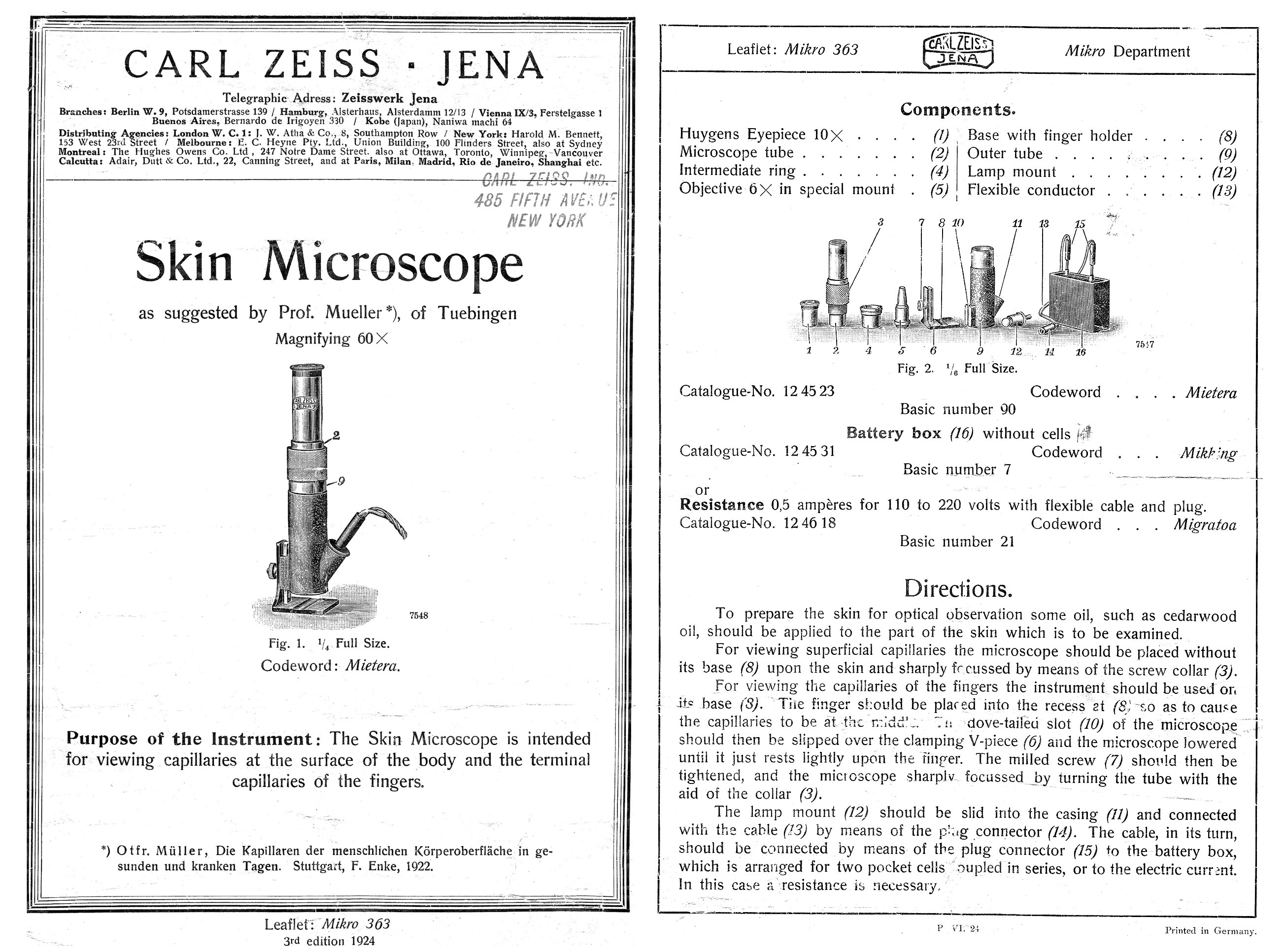
Starting about 1916, Professor Otfried Muller of Tubingen, Germany studied many types of
capillaries including those of the nail fold; this culminated in his magnificent 1922 book and color atlas
'Capillaries of the Surface of the Human Body in Health and Disease.' Zeiss credited him with the design
of the microscope shown here in the engraving from Muller's book from 1922, and also above.
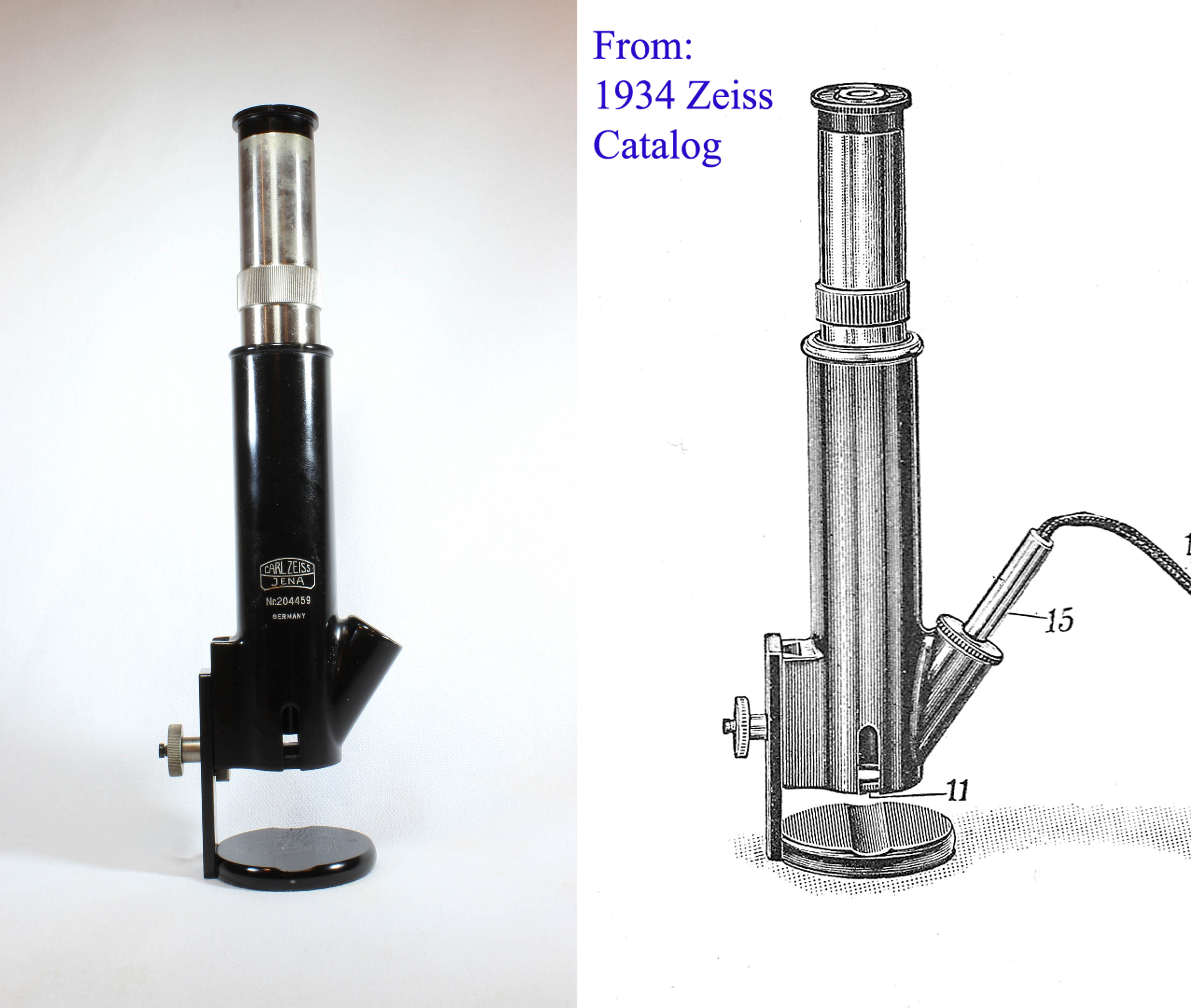
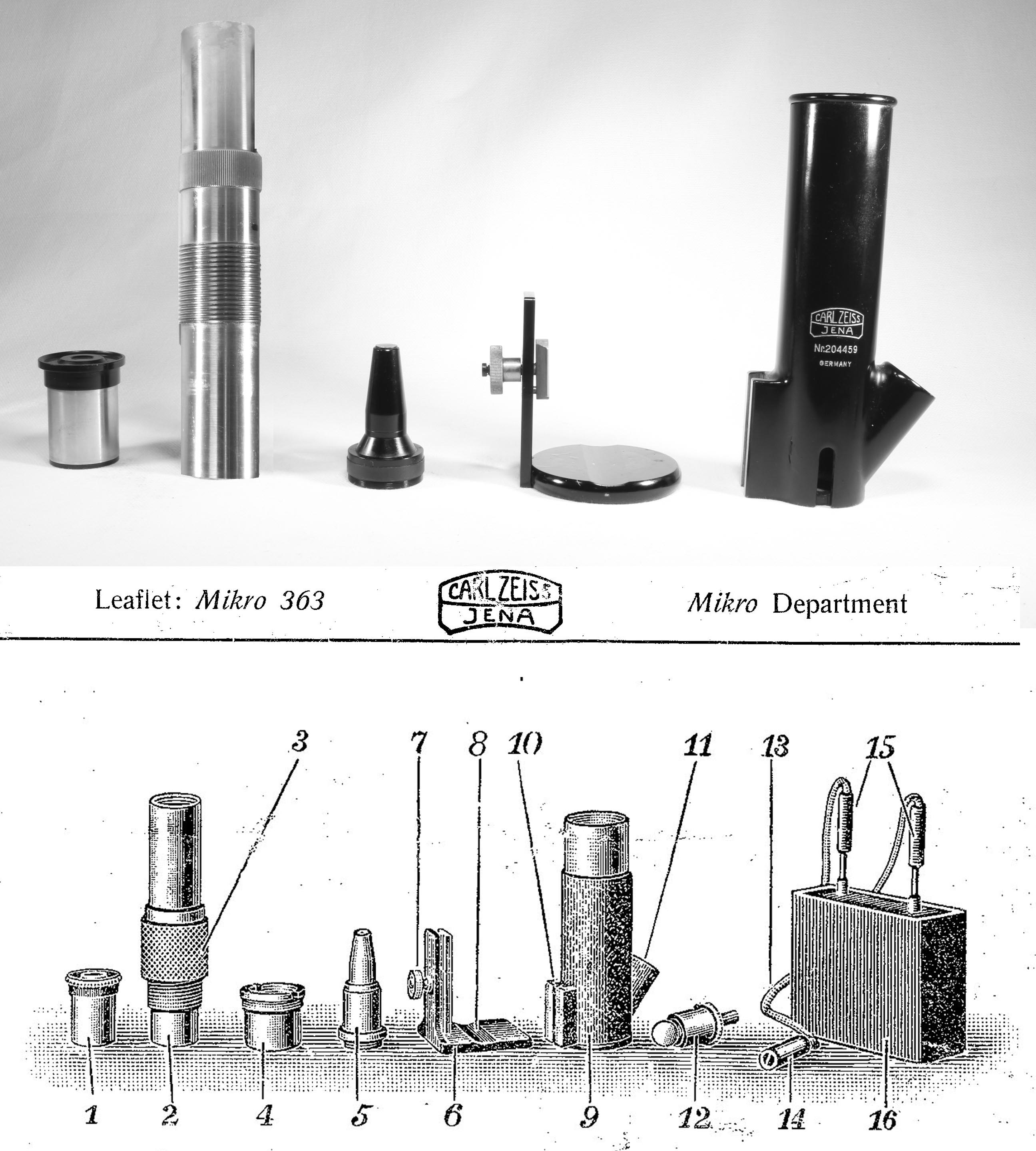
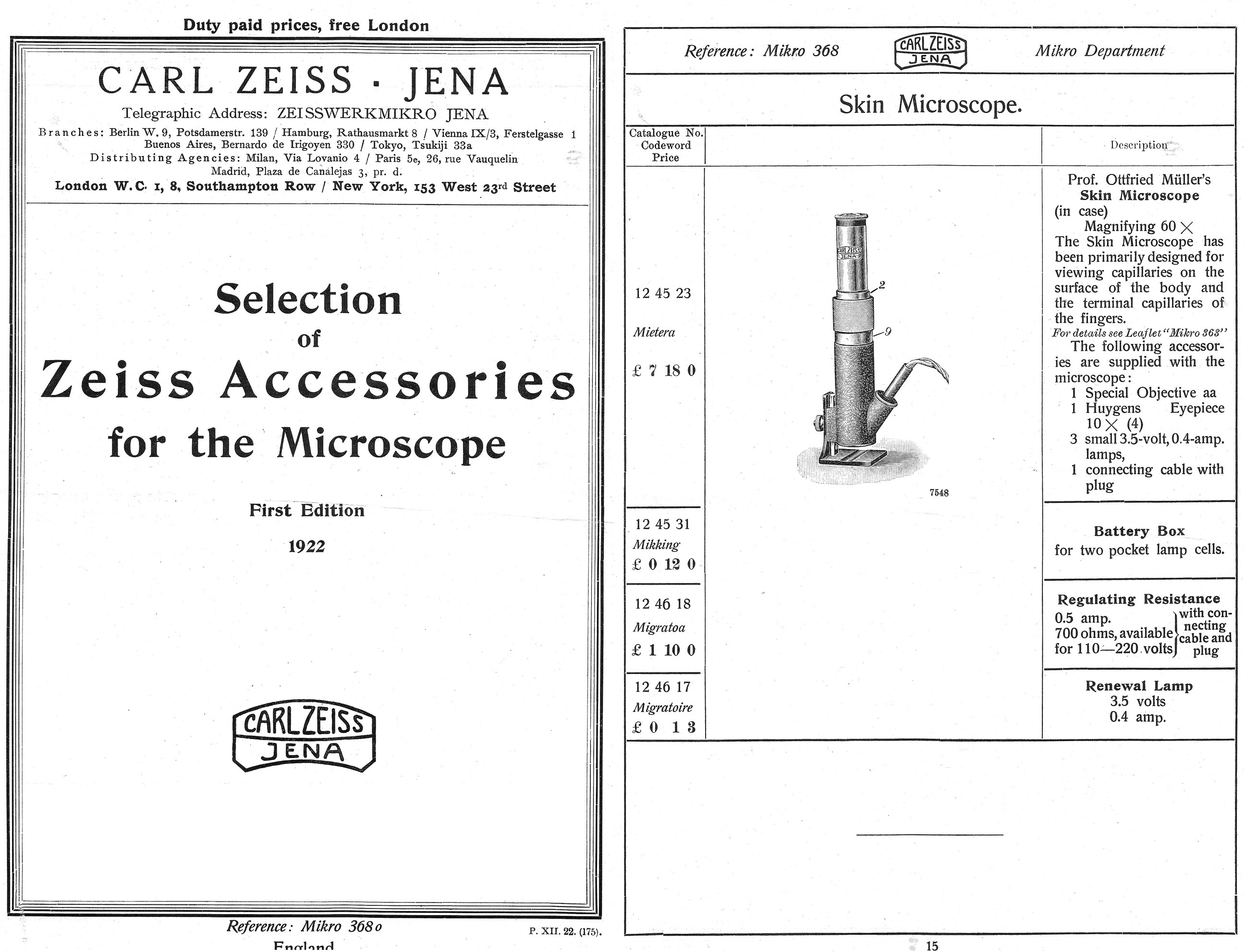
The design of this microscope changed slightly over the years. As shown in the accompanying
1924 instruction
leaflet, and also in the
Zeiss 1924
catalog of accessories, it originally had a square shaped base, but by the date the above example was
produced (1928) it had a round foot, as pictured
in the 1934 Zeiss catalog. Also, by the time this instrument was
produced, the 'intermediate ring' was not part of the instrument; the optical tube screwed into the
'outer tube' directly. Another change was a transition to the use of a standard 10X eyepiece,
as opposed to the original design pictured in Muller's illustration to the left, which shows
the upper optical elements integrated into the tube itself.
Other manufacturers made nailfold capillary microscopes;
Bausch and Lomb marketed one for this purpose about 1929. From the early 1920's until at least 1934,
Muller's Skin Microscope was featured in their catalogs. By 1934 Zeiss offered a
new model of skin microscope, although the 'Small Muller Capillary Microscope' was still offered.
This newer skin microscope could also be adapted to the study of other skin capillaries such as those around
the lips, and could be supplied with apparatus for photographing the capillaries.
Today the technique of capillary microscopy is occasionally used to help diagnose rheumatic diseases,
especially scleroderma and other connective tissue diseases. The normal capillaries of the skin at the base of
the nail are relatively uniform in thickness and often (but not always) relatively straight curved loops
resembling the closed end of a 'bobby pin'; in these diseases however they become deformed both in course and
diameter and there may also be associated tiny hemorrhages or avascular areas lacking capillaries completely.
These features are only visible with magnification greater than that provided by, for example, a
magnifying glass. An ordinary microscope could be used for this purpose with top-side illumination,
but having the subject's finger hold steady on a stage without support for his or her arm, would be very
difficult. This is the main advantage of this kind of microscope, as the depression in the foot plate keeps
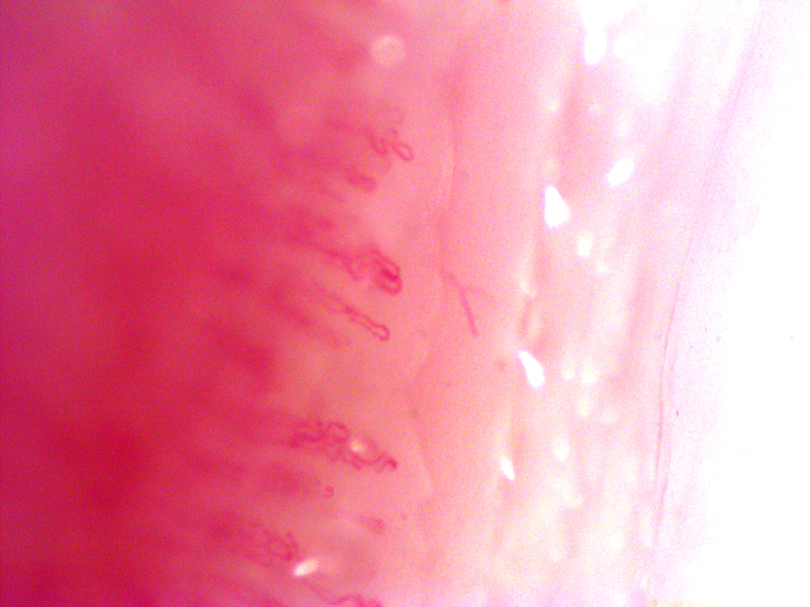
the finger centered, and the subject's arm can rest on the table. It has been said that tortuosity of the
vessels can also reflect these conditions, but as seen in the illustration here, from a normal male subject
(the author!), tortuous nail fold capillaries can be found in normal persons, especially males.
One of the difficulties in using the instrument shown here is a narrow depth of focus, hence the instrument
must be focused up and down to visualize the capillaries across the field of view.
This is a marked disadvantage, especially in photography or for quantitative measurements.
A recent form of nailfold capillary microscope with integral digital camera, has a mechanical stage
which gently grips the finger, and this makes positioning and imaging a bit easier.