MICROSCOPE-ANTIQUES.COM © 2013-15.
MODEL: SEIBERT 'LARGE NO. 2' MICROSCOPE
Same model as Koch used to study Anthrax
DATE: c. 1890
MAKER: SEIBERT, GERMANY
MODEL: 'LARGE NO. 2'
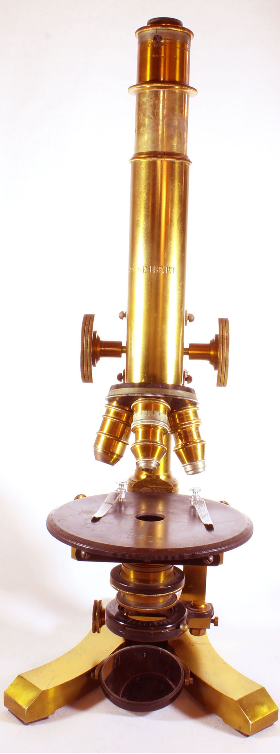
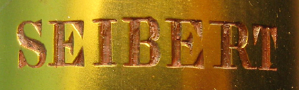
SIGNED: 'SEIBERT'
SERIAL NUMBER: 7880
PLEASE CLICK ON THE PICTURES FOR ENLARGED IMAGES
DESCRIPTION:
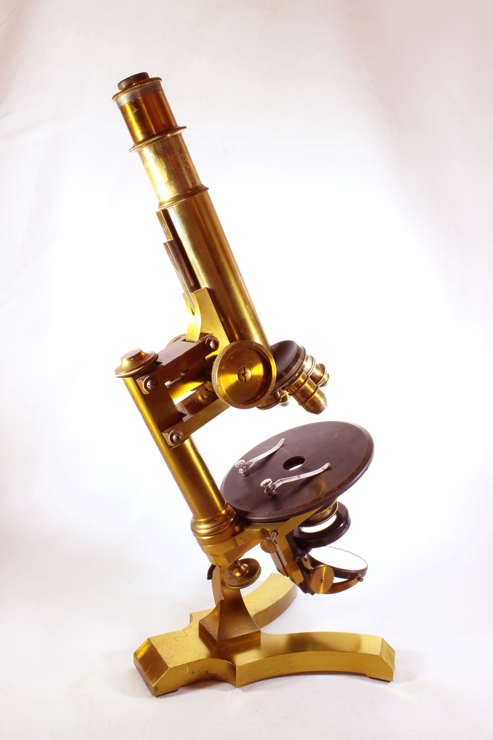
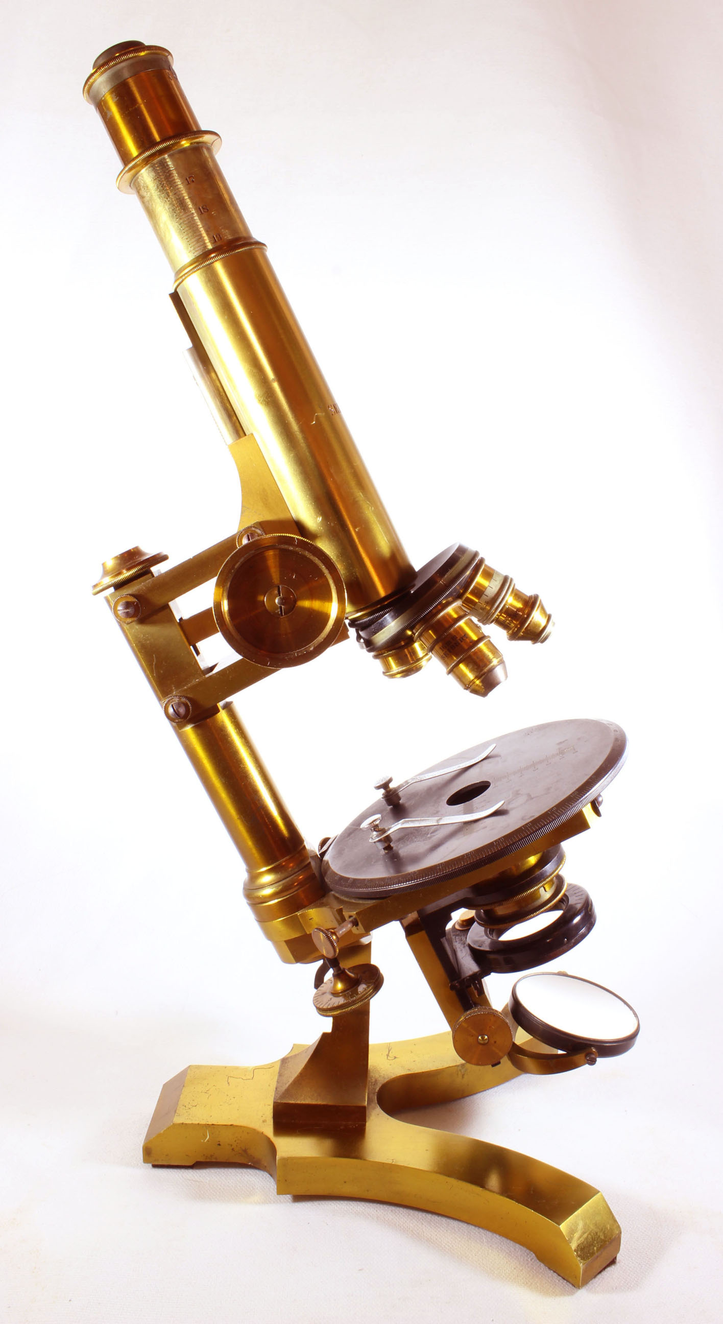
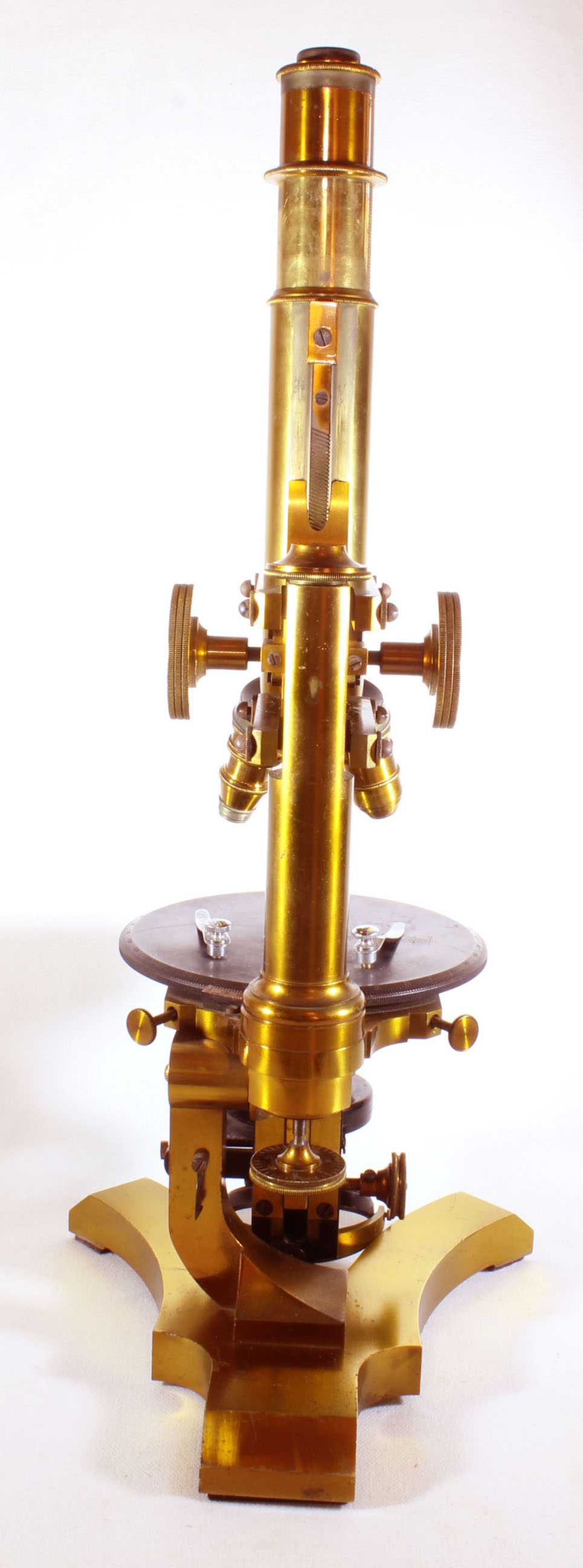
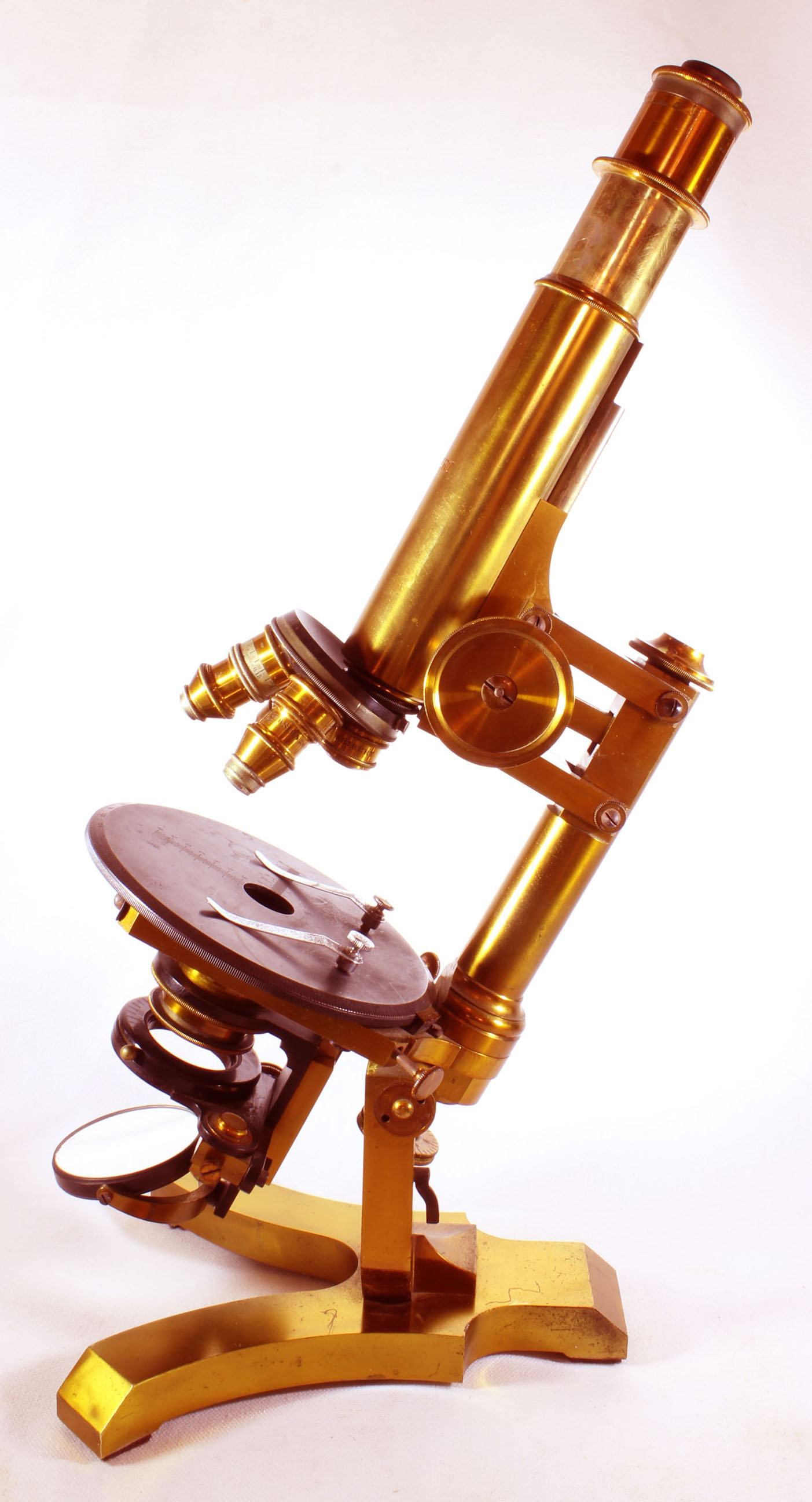
 This microscope is a fine example of a large Seibert Microscope, the No.2 Model. It arises
from a V-shaped claw foot, on a single C-shaped pillar curving to the left, leaving convenient access to the fine focus knob from the
right side. The gimballed mirror is plano-concave and swivels but is permanently fixed to the end of the tailpiece. The tailpiece is fixed and does not 'swing.'
There is an oxidized black indicator-pointer for registering the graduations on the fine focus; this is attached to the C-shaped pillar. The indicator
is rotated into position when the microscope is vertical; in the inclined positions it is not usable and is swung out of the way.
This microscope is a fine example of a large Seibert Microscope, the No.2 Model. It arises
from a V-shaped claw foot, on a single C-shaped pillar curving to the left, leaving convenient access to the fine focus knob from the
right side. The gimballed mirror is plano-concave and swivels but is permanently fixed to the end of the tailpiece. The tailpiece is fixed and does not 'swing.'
There is an oxidized black indicator-pointer for registering the graduations on the fine focus; this is attached to the C-shaped pillar. The indicator
is rotated into position when the microscope is vertical; in the inclined positions it is not usable and is swung out of the way.
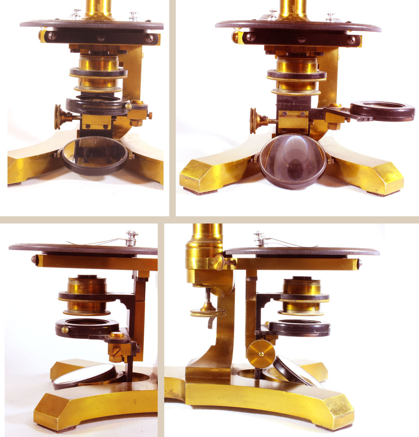
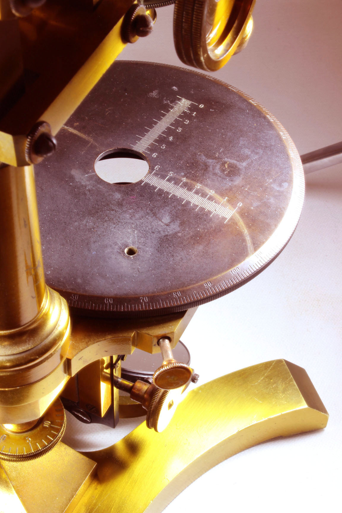
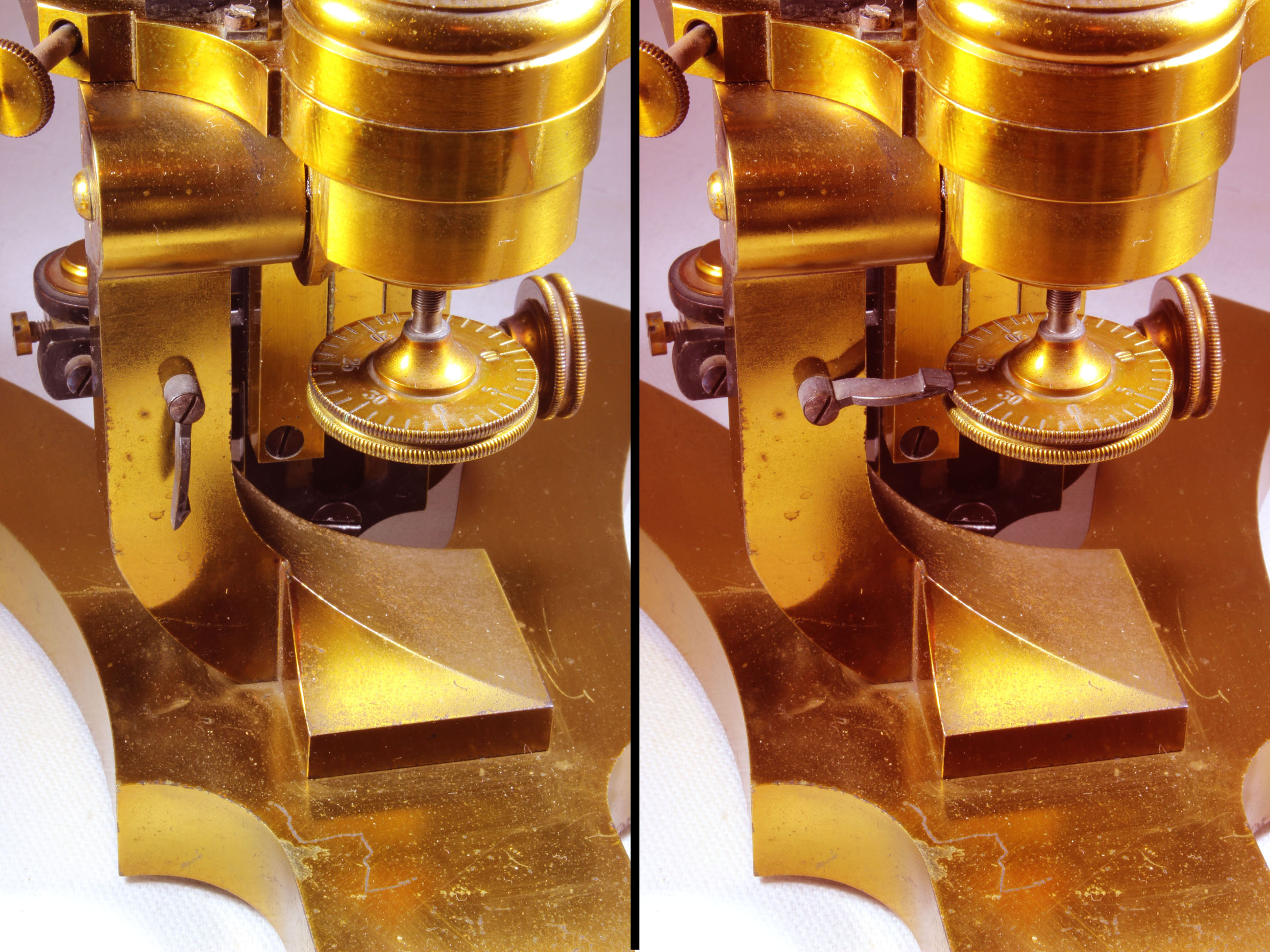
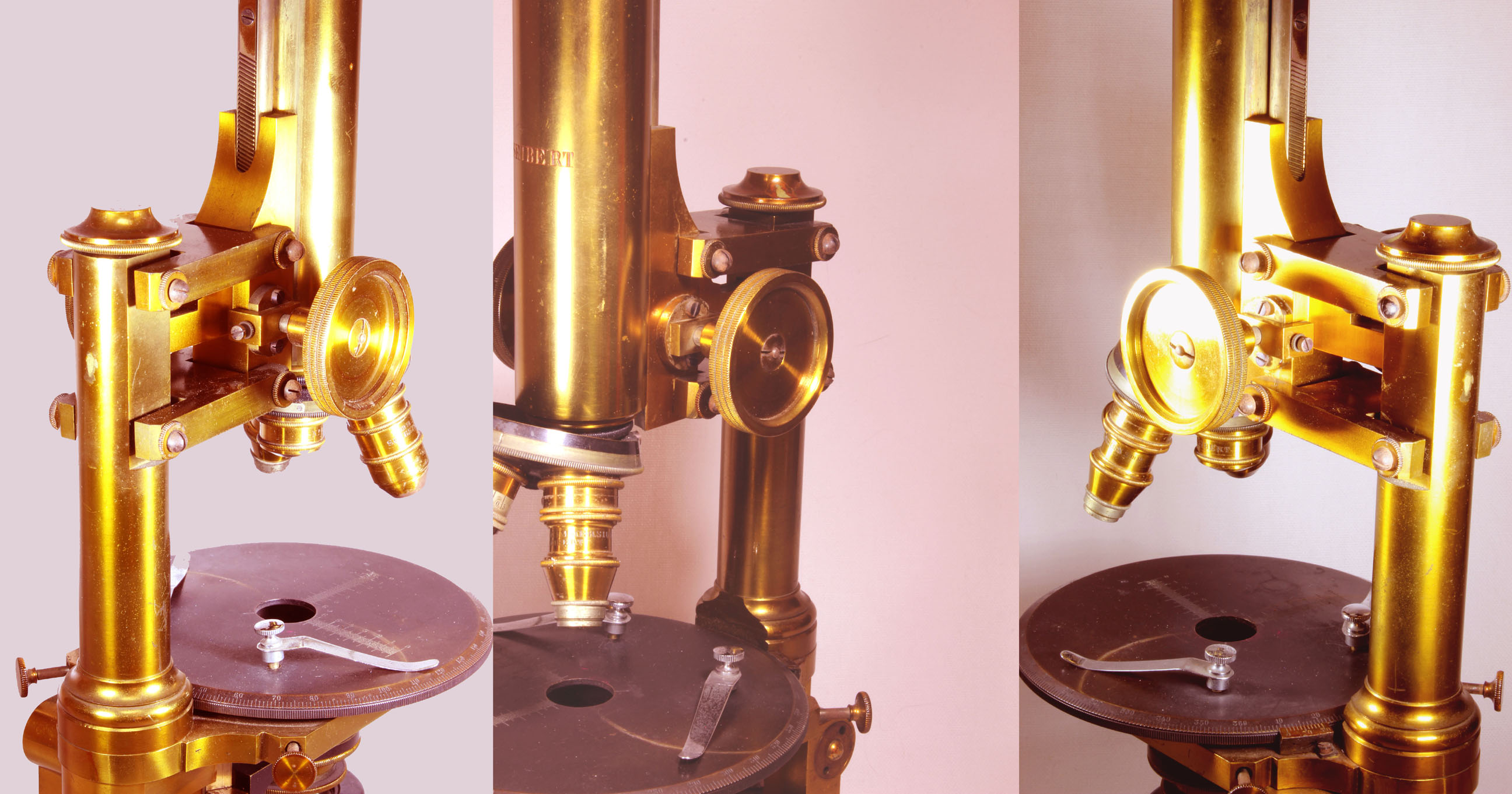
The substage, pictured in detail above, is focused by diagonal rack and pinion, carries two separate
appliances. The lower one is an iris diaphragm, the upper holds the separate condenser. The iris assembly can swing out of the optical axis after
releasing a sprung catch. There is a Abbe condenser, but in the box an additional fitting can replace it to hold one of the included three stops.
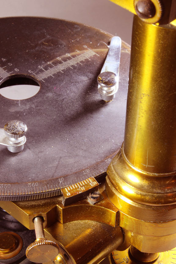
The stage is of oxidized brass and has two stage clips. It rotates and is calibrated around its outside bevelled edge in 1 degree increments and there is a lacquered brass
vernier to further refine the readings of stage rotation to a tenth of a degree. There are two knobs for adjusting the centration of the stage.
In addition, on the stage are two sets of graduations, arranged ninety degrees
from each other to act as a finder to locate a specific location on a slide. Two removable nickel or chrome-plated stage clips are present.
 The coarse focus is by diagonal or helical rack and pinion and the casing for the pinion has screw adjustments.
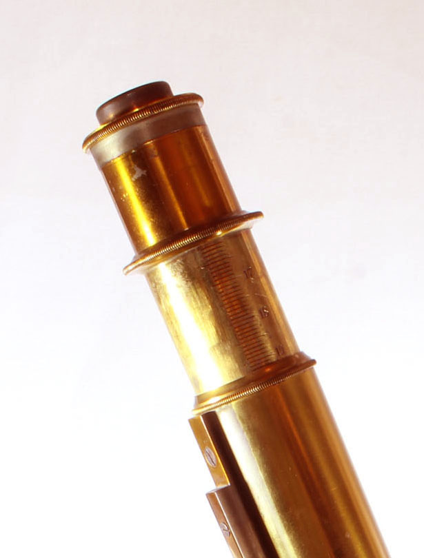
The instrument has an inclination joint. The body tube is signed in block letters, 'SEIBERT.' There is a graduated draw tube and a quadruple
nosepiece with four original Seibert objectives including one with a correction collar.
The objectives are labelled:
- 'No. 0, SEIBERT
- '16 mm, SEIBERT'
- '4 mm, SEIBERT' (with correction collar)
- 'Homogen Immersion 2 mm, SEIBERT'
It is in fine operating condition with a few thin scratches on the body tube and foot. There are a few small lacquer losses, but 99% or more remains.
There is only a single eyepiece with crosshairs. The eyepiece has a screw on its side which
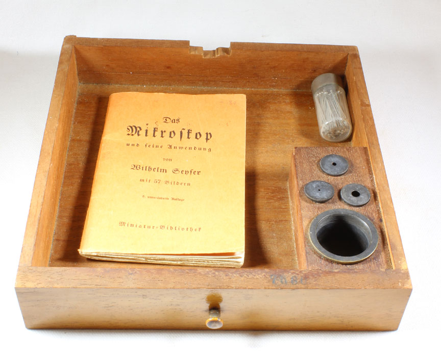
fits into a matching slot on the drawtube. The original upright case is with microscope with lock but no key; it has the original drawer above the microscope which houses the aperture
stops and their holder. An interesting little booklet, written in German, accompanied the microscope entitled:
Das Mikroskop, und feine anwendung , von Wilhelm Genfer, mit 57 Bildern, c. unveranderfe auflage , Minatur Bibliothe
(The Microscope and its application by Wilhelm Genfer, with 57 illustrations ---?--- edition, Miniature Libraries-note that in those years the letter 's' was written as an 'f')
HISTORY OF THE SEIBERT MICROSCOPES.
 The famous Carl Kellner's
Optical business was taken over by Belthle and Rexroth in Wetzlar Germany. Ernst Gundlach, along with Wilhelm
Seibert and Heinrich Seibert, worked for Kellner in the 1850's. In 1859 Gundlach set up his own workshop, taking
with him the Seibert brothers as employees. As was to be the case many times in future endeavors, Gundlach had
poor business skills and left the company in debt, fleeing to England about 1860. In 1865 he returned from
England, this time setting up a business in Berlin. Again the Seiberts worked for him from about 1866.
By August of 1872, the business was again in financial trouble, and
The Seiberts, along with the financial help of Georg Krafft, a businessman, bought the foreclosed business
(and its debts!). Because Gundlach had to agree not to found a business in Germany, he emigrated to the United
States. Meanwhile, Seiberts and Krafft moved back to Wetzlar. The company was known as Seibert and Krafft from
1871-1884, and and W. and H. Seibert from 1884 to about 1925.
The famous Carl Kellner's
Optical business was taken over by Belthle and Rexroth in Wetzlar Germany. Ernst Gundlach, along with Wilhelm
Seibert and Heinrich Seibert, worked for Kellner in the 1850's. In 1859 Gundlach set up his own workshop, taking
with him the Seibert brothers as employees. As was to be the case many times in future endeavors, Gundlach had
poor business skills and left the company in debt, fleeing to England about 1860. In 1865 he returned from
England, this time setting up a business in Berlin. Again the Seiberts worked for him from about 1866.
By August of 1872, the business was again in financial trouble, and
The Seiberts, along with the financial help of Georg Krafft, a businessman, bought the foreclosed business
(and its debts!). Because Gundlach had to agree not to found a business in Germany, he emigrated to the United
States. Meanwhile, Seiberts and Krafft moved back to Wetzlar. The company was known as Seibert and Krafft from
1871-1884, and and W. and H. Seibert from 1884 to about 1925.
 This was the second largest of Seibert microscopes at the time it was produced.
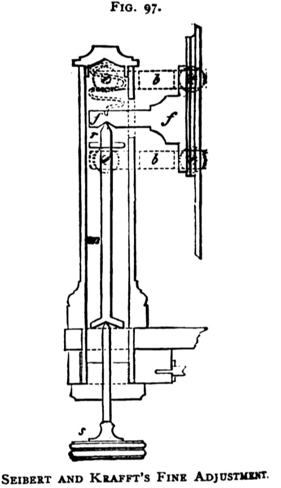
It features a solid parallel linkage fine focus mechanism controlled 'without friction' from beneath the stage. This
was illustrated and described in the Journal of the Society of Arts (October 1, 1886)
as shown in the figure reproduced to the right. According to a citation in the Transactions of the
Journal of the Royal Microscopical Society, the Seiberts introduced this mechanism in 1876, but Gundlach microscopes which date to earlier contain this mechanism; importantly, the Seiberts were making microscopes for Gundlach prior to going into
business on their own; clearly this mechanism is from before 1869, since a
Gundlach microscope from that year is
equipped with a parallel linkage fine focus controlled from below. Timo Mappes has provided evidence (in a quotation from Seibert), that Gundlach came up with the idea for this focusing mechanism after seeing a similar control on a balance or scale invented by the French mathematician de Roberval; although this idea may have originated with Gundlach, (and de Roberval), the Seibert company continued its use for decades.
This was the second largest of Seibert microscopes at the time it was produced.
It features a solid parallel linkage fine focus mechanism controlled 'without friction' from beneath the stage. This
was illustrated and described in the Journal of the Society of Arts (October 1, 1886)
as shown in the figure reproduced to the right. According to a citation in the Transactions of the
Journal of the Royal Microscopical Society, the Seiberts introduced this mechanism in 1876, but Gundlach microscopes which date to earlier contain this mechanism; importantly, the Seiberts were making microscopes for Gundlach prior to going into
business on their own; clearly this mechanism is from before 1869, since a
Gundlach microscope from that year is
equipped with a parallel linkage fine focus controlled from below. Timo Mappes has provided evidence (in a quotation from Seibert), that Gundlach came up with the idea for this focusing mechanism after seeing a similar control on a balance or scale invented by the French mathematician de Roberval; although this idea may have originated with Gundlach, (and de Roberval), the Seibert company continued its use for decades.
According to the 1893 edition of 'The Microscope' by Dr Henri Van Heurck, Seibert manufactured:
excellent microscopes, the mechanical part being as carefully constructed as the optical,
leaves nothing whatever to be desired...Stand No. 2 though of smaller proportions, is almost identical with
the last
(No. 1).
According to the description of the number 2 in Van Heurck, it had:
...two movements for focusing; the rapid movement by rack and pinion, and the slow movementby
micrometrical screw, the of which is graduated. There is a draw tube, divided by a scale. The stage is rotary,
and the illuminating apparatus consists of an Abbe condenser, furnished with an Iris diaphragm. The different
pieces of apparatus which accompany it consit of a nose-piece for four objectives, a moveable eyepiece micrometer,
polarizing apparatus with graduated circle, camera lucida, bulls-eye on stand, ordinary objectives
Nos. 0 to VII, an homogenous immersion objective 1.12 inch with 1.3 numerical aperture, and
simple oculars Nos. 0 and I, and periscopic oculars Nos. II and III giving magnifications from 18 to 1500
diameters. The instrument enclosed in a strong box, with test objects and other accessories such as glass
slips and cover glasses is sold for £46 10s.
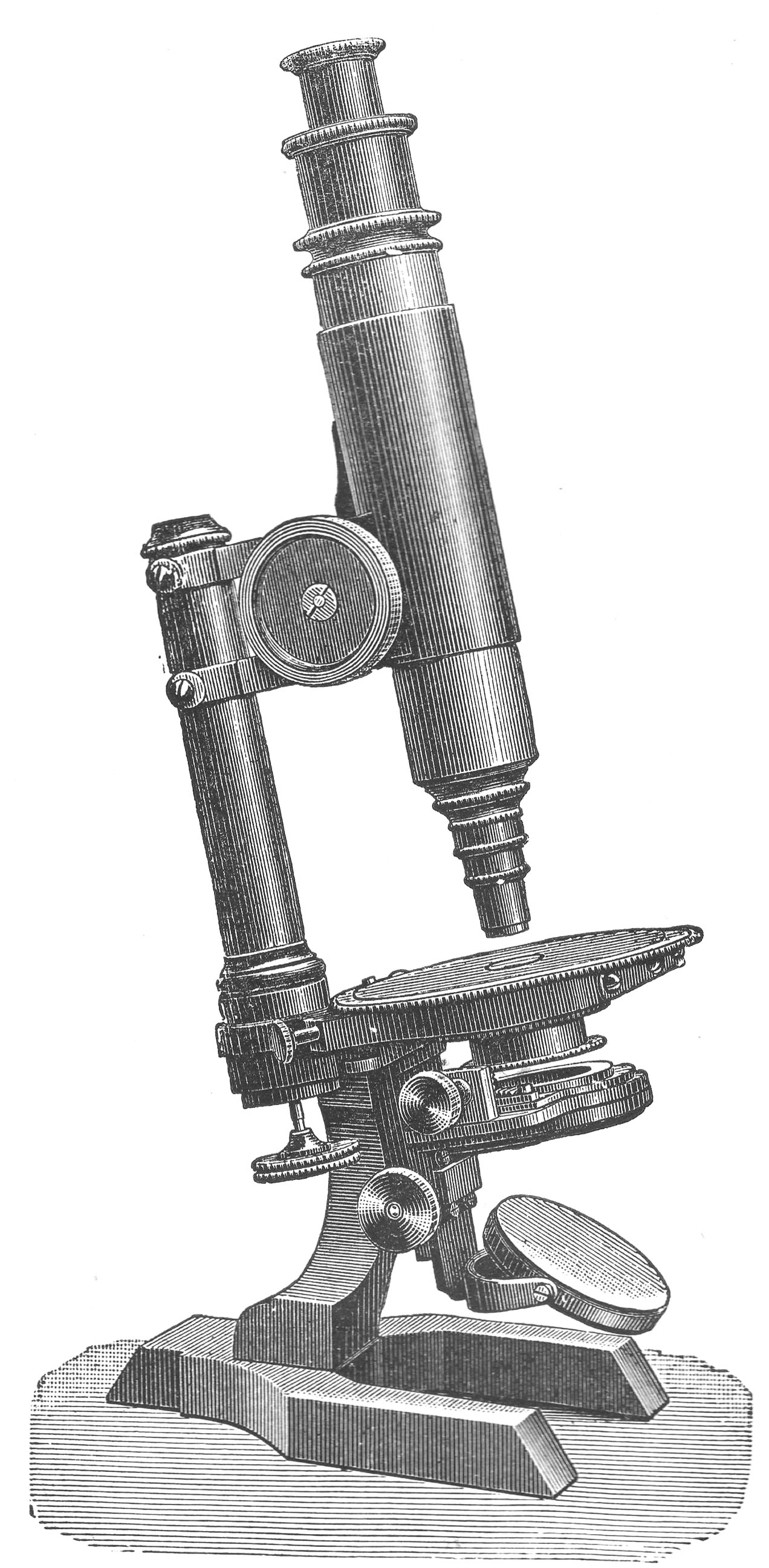
Note that the microscope pictured in the 1893
Van Heurck (shown to the left) had changed slightly from the model pictured on this web page, and indeed the microscope featured on this webpage
was a transition from an earlier model; for a comparison of this microscope with one before and one dated after this one, here.
Gundlach microscopes made in Berlin in the late 1860's have many similar features to the later microscope pictured here above.
These features include the C-shaped pillar, the fine focus knob location, and a parallel linkage fine focus mechanism.
Indeed the parallel linkage fine focus, first devised by de Roberval for his Balance, and first conceived of for use on a microscope by Gundlach, was part of the design not only of some many Seibert microscopes, but also Leitz, Zeiss and others.
In fact, a small microscope utilizing this type of fine focus (but with the control knob on top) by Leitz is in this collection. It was even used (with the control knob at the top instead of the bottom of the pillar), by the American maker Walter Bulloch on the Joint effort with a Professor Bastin (the so-called Bastin-Bulloch microscope).
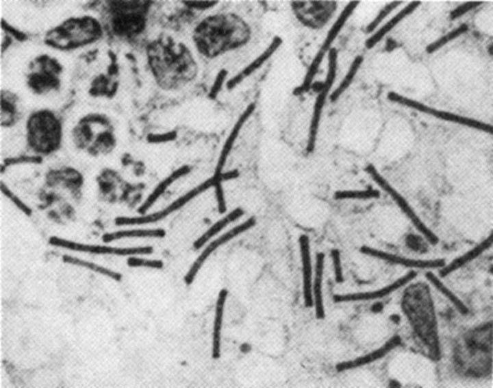 Of great historical interest, this model, but thirteen years earlier, was apparently used by Robert Koch when he worked on anthrax. Koch was the first physician to use oil immersion objectives, and was the first to publish a photomicrograph of the Anthrax bacillus(left).