CALCITE CRYSTAL TO DEMONSTRATE DOUBLE REFRACTION
MAKER: ANDREW ROSS
c. 1847
'DOUBLE IMAGE PRISM'
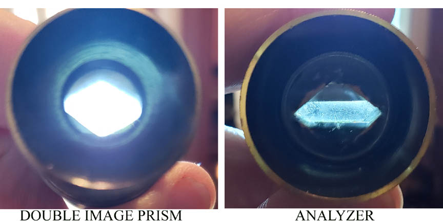 DESCRIPTION: Among the apparatus supplied with 19th century microscopes, an interesing accessory is the eyecap containing a crystal of calcite, called the 'double image prism'. Superficially, this resembles the 'analyzer' eyecap to be used with the substage polarizer for viewing anisotropic minerals and other birefringent materials under polarized light, often to spectacular effect. This eyecap however is different. Unlike the Nicol prism analyzer, it contains a single cut crystal. If one looks at an object through this eyecap,(with or without a lens), a double image is seen. Hence the name 'double image prism'. In use, this eyecap is used to demonstrate birefringence under the microscope, making use of point sources of ordinary or polarized light. Various experiments can be carried out to illustrate the behavior of light passing through the calcite (see below). This type of eyecap can be differentiated from the analyzer eyecap in that when the cap is viewed in front of a faint light or faintly lighted object, the opening in the prism appears much wider than the narrow appearing almost slit-like opening of analyzer eyecaps. The Smith & Beck Best No 1 microscope of circa 1860 in this collection also came with apparatus for double image prism experiments.
HISTORY OF THE CONCEPT OF DOUBLE REFRACTION AND ITS DEMONSTRATION:
The appearance of a double image when viewed through a piece of calcite crystal was first noted by Erasmus Bartholinus in 1669. He did not understand the nature of the images or the concept yet to be described, that is that the two images were each composed of polarized of light, with the axis of polarization oriented 90 degrees to each other. In 1690 Christian Huygens realized that the two images could be extinguished by another crystal with its optical properties oriented 45 degrees from the first, and concluded that waves of light were cancelling each other out. Although he did not understand the direction of these waves, he was apparently the first to realize that the two different images were polarized. In 1815 Sir David Brewster discovered the law giving the relationship between the refractive index and the angle of incidence of a light beam at which the beam is totally polarized. In 1828 William Nicol of Scotland invented the Nicol prism which uses two pieces of calcite to allow only one beam of polarized light oriented in a single direction, to pass through it. This was the first practical polarizing filter and led to many important advances. Finally, in 1834, Henry Fox Talbot adopted polarization apparatus to the microscope and started the study of mineral thin sections, the field of geology using the petrographic microscope for identification of minerals.
Although much of the use of the microscope in the 19th century was recreational, reproduction of some experiments were felt to be important. Experiments using the 'double image prism' eyecap to illustrate the properties of birefringence and polarized light became popular after they were described by Legg in the Transactions of the Microscopic Society of London, Vol 1 of December 1846. These experiments were illustrated in some detail and continued to be popular, as exemplified by the fact that their description was repeated in even greater detail twenty years later in Richard Beck's Treatise on the Construction, Proper Use and Capabilities of Smith, Beck, and Beck's Achromatic Microscopes of 1865, along with illustrations of the apparatus itself. Of great interest, the apparatus illustrated by Beck was essentially identical to the one supplied with the Ross Bar Limb Microscope No 324. This suggests to me that one firm actually supplied the selenite condenser to both firms, perhaps Darker himself.
THE EXPERIMENTS:
If, while using ordinary substage illumination, one uses the 'double image prism' in place of the ordinary eyecap, while looking at a small hole in an opaque slide made of e.g. brass, one will see the dot of light from the hole and a second dot near it. If one rotates the eyecap, the dots will appear to circle each other.
Shown here is a series of images captured from the Ross Bar Limb microscope using a polarizer. If a polarizer is used below the perforated slide and the experiment repeated, the rotation will still occur, but each dot will alternately disappear as the location at which extinction is reached for each and reappear as the rotation is continued, cycling at 45 degree intervals of rotation.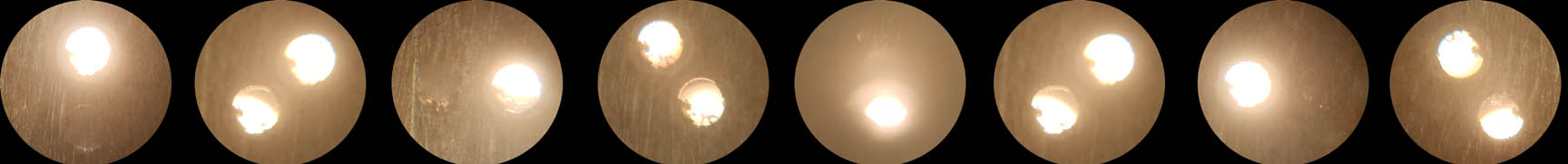
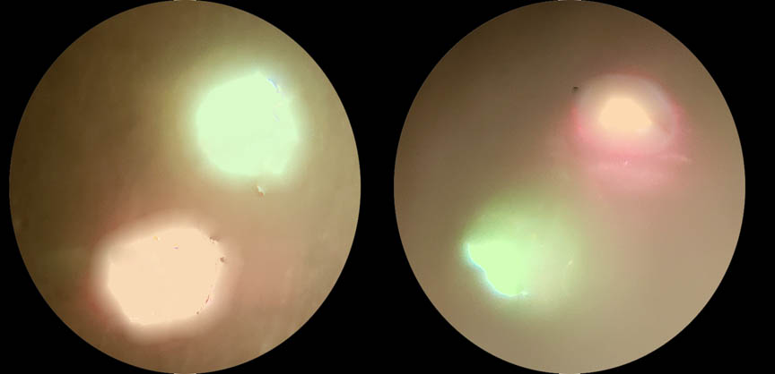 For even more effect, a wave plate retarder, such as a quarter wave plate or selenite placed below the perforated slide will, in addition, show the dots alternately changing to opposing colors as the eyecap is rotated. The image to the left shows that as the double image prism is rotated 180 degrees, the two dots change color as their wave vectors are oriented 90 degrees from each other. Several additional experiments are also described in the works cited above, but some require additional accessories such as a second calcite 'double image prism'. These experiments do not require a microscope these days, as one can simply look through the eyecap without a microscope (or even an eyepiece), at the hole in the brass aimed at an ordinary light source, rotating the cap as described above. If one uses a white screen on a computer as the light source, since the white light from the computer screen is polarized, the experiments with a polarized light source, with or without a selenite or retarder, are easily reproduced. Actually performing these experiments gives one a hands-on understanding of the principals involved.