| DESCRIPTION | HISTORY |
Please Click On Any Picture for a Larger Version
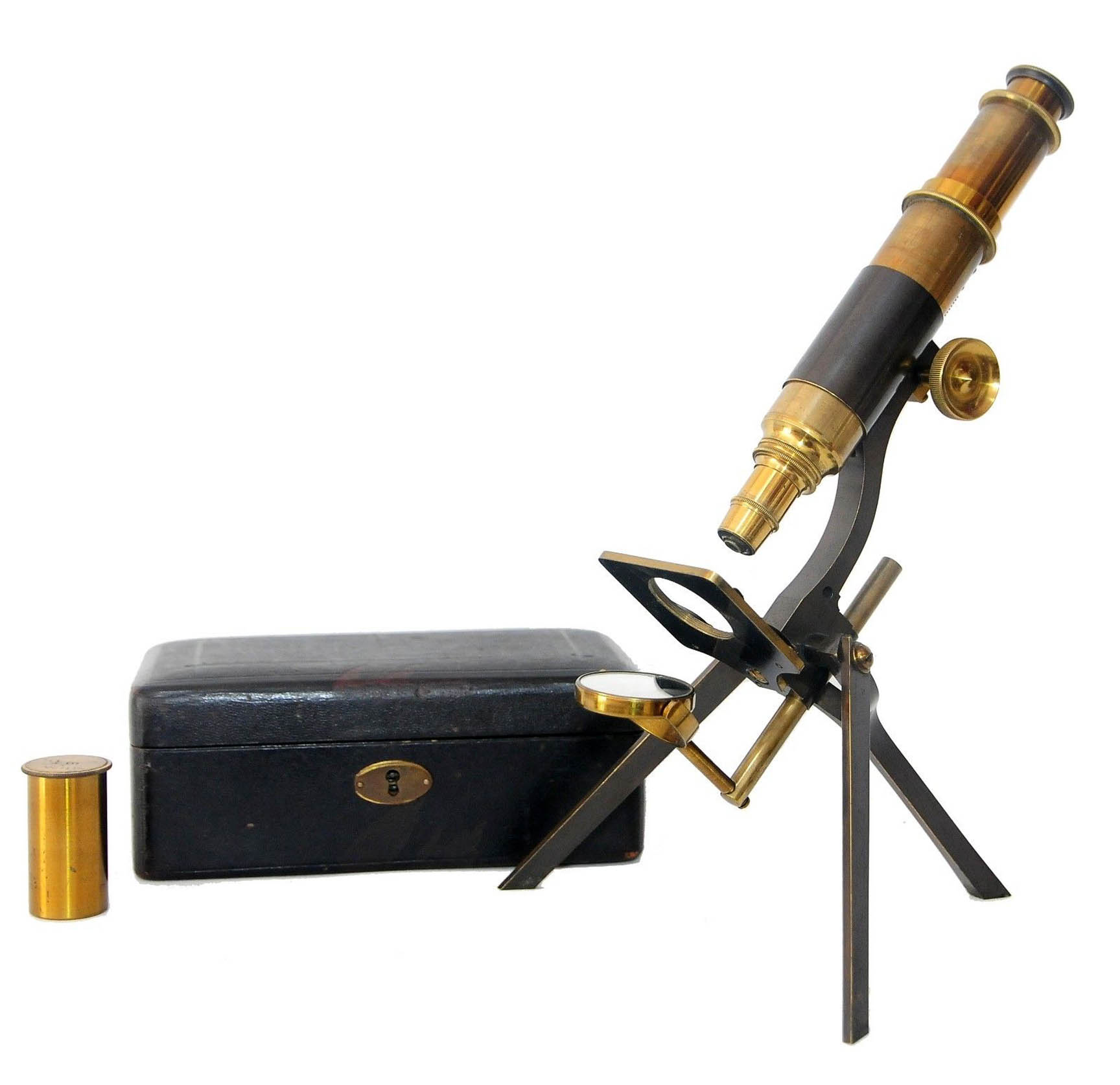
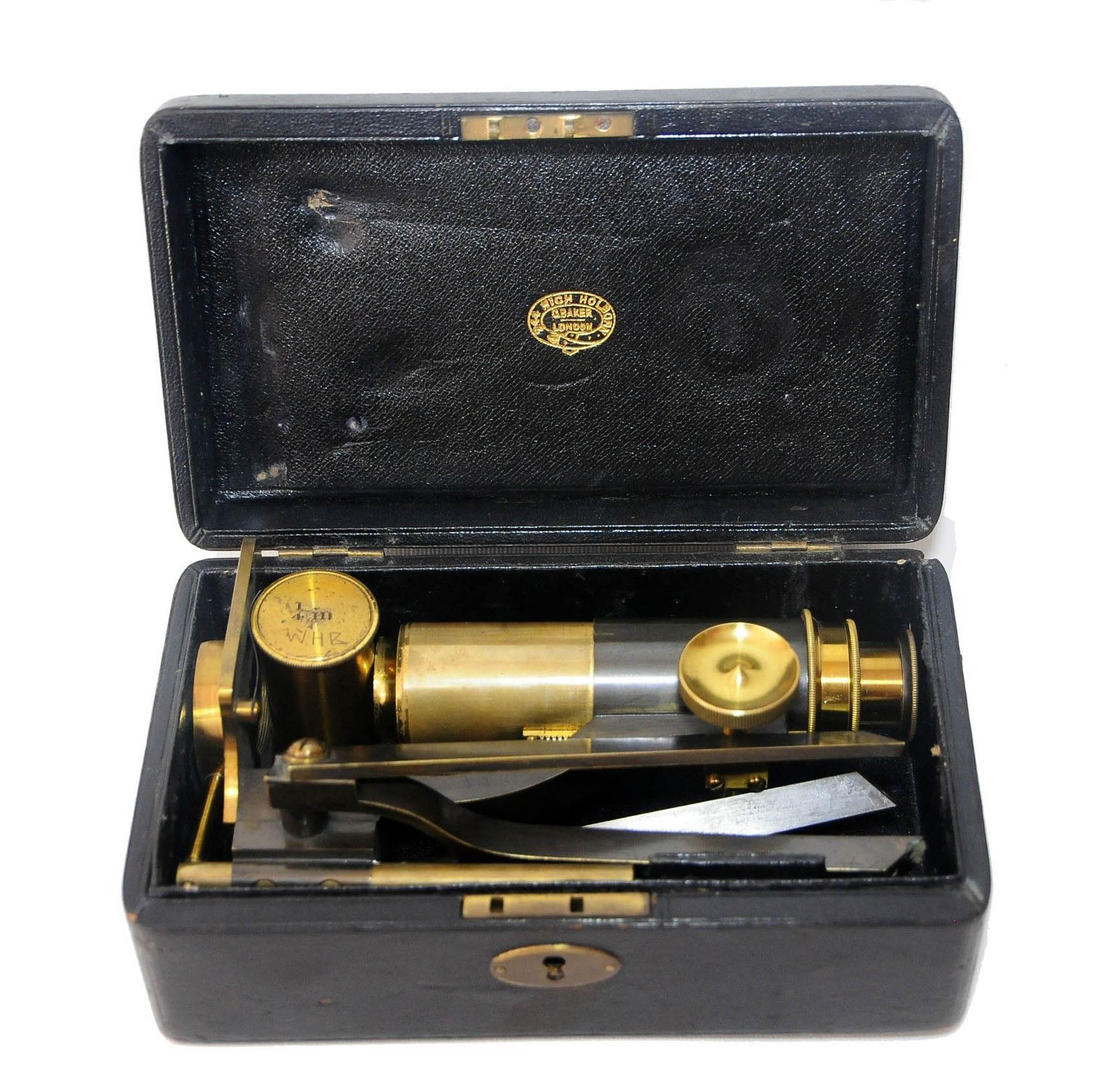
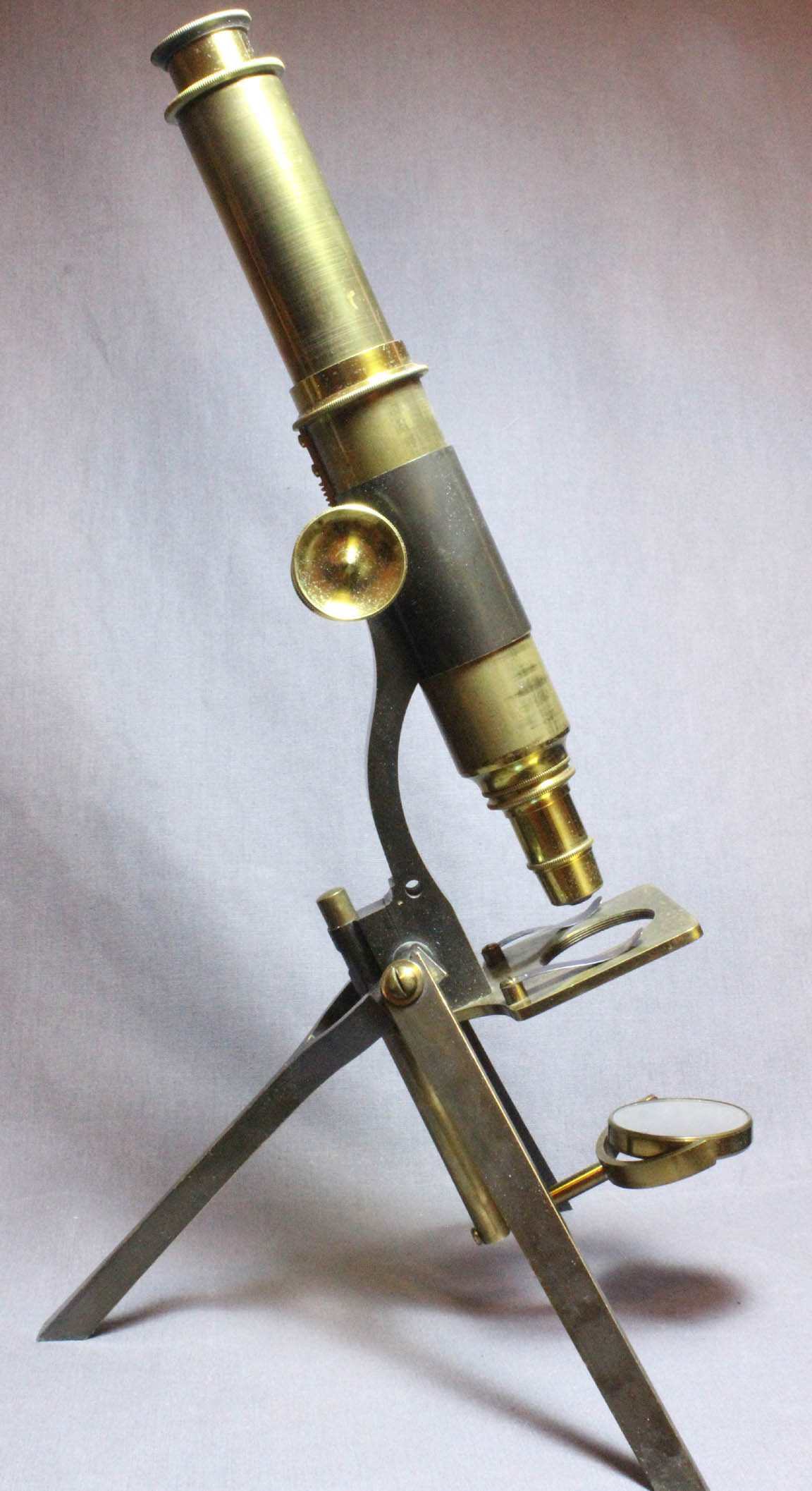
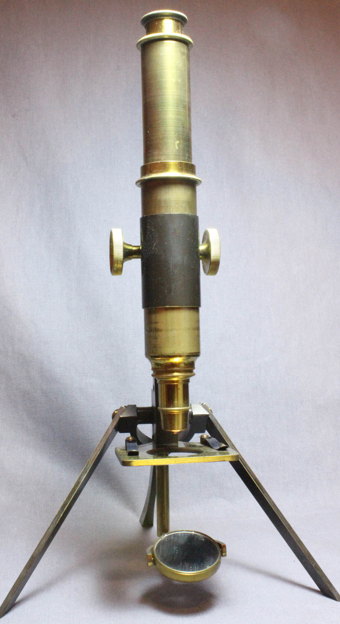
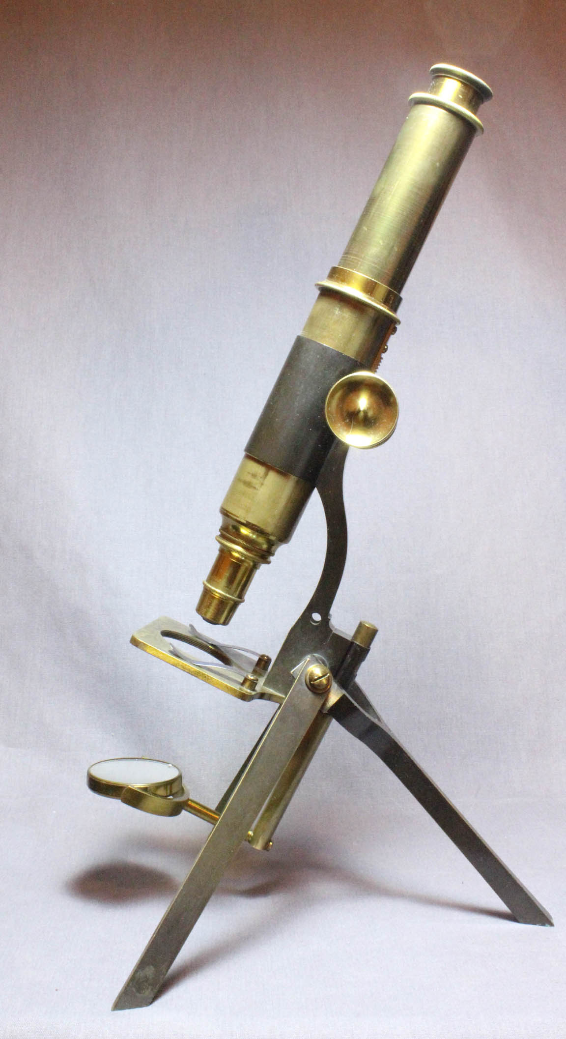
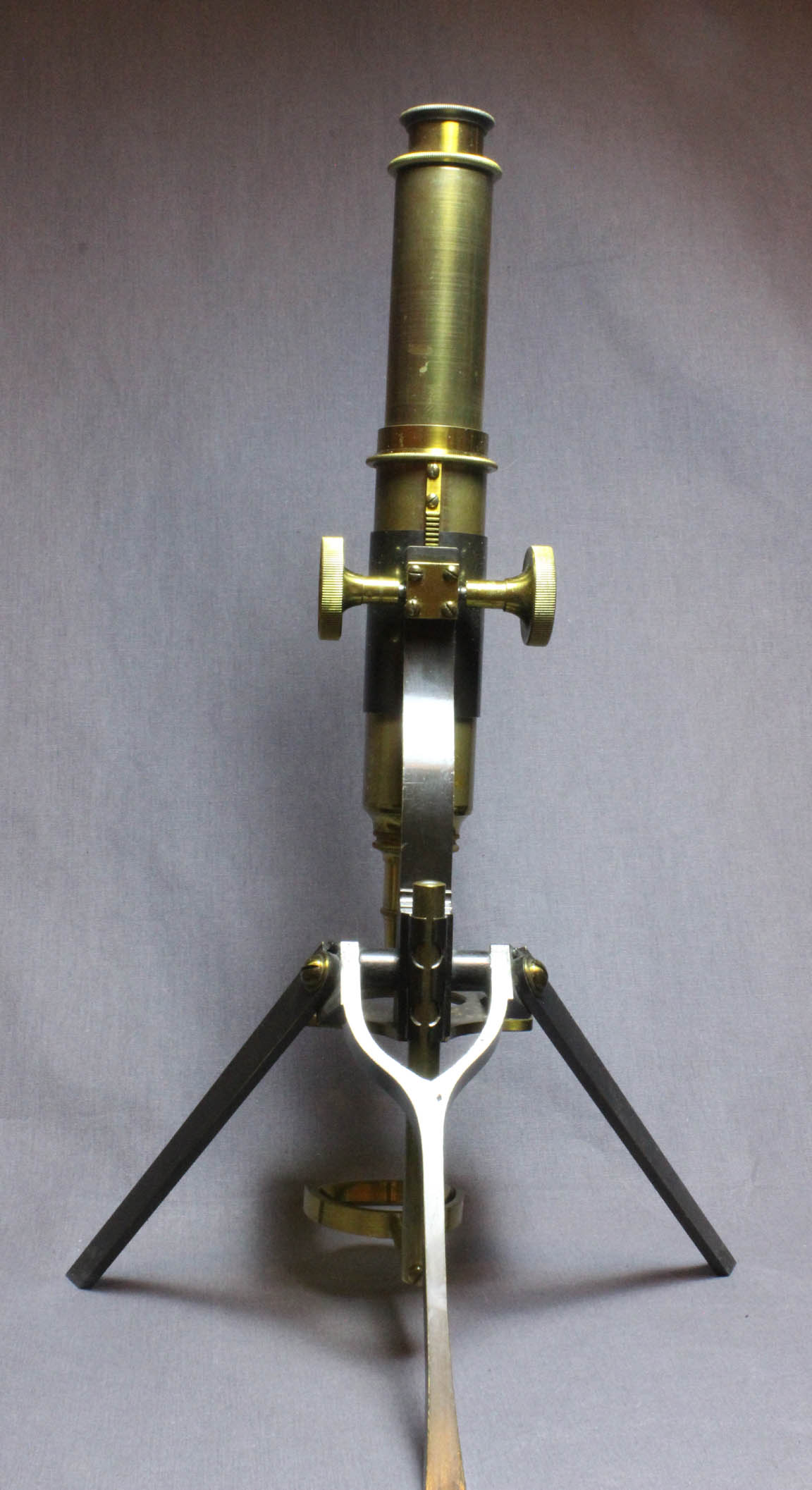
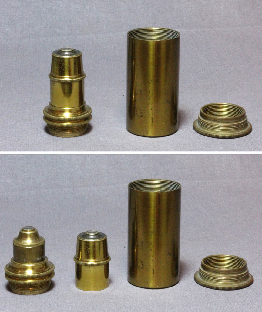
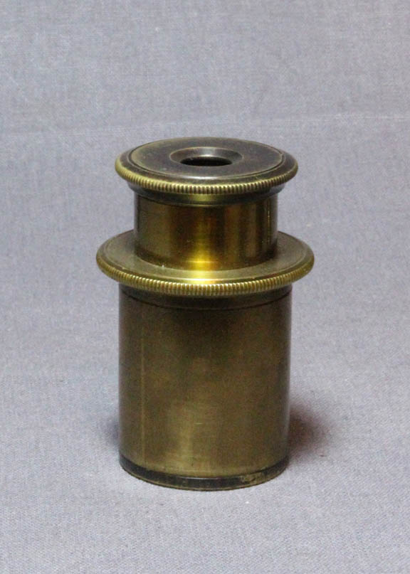 This microscope is supported by a folding tripod foot. The rear leg is Y-shaped. All three legs have a rectangular profile. The plano-concave mirror rides on a rod which can be pushed up or pulled down. The rectangular stage has two slide clips and the central opening is threaded. Coarse focus is by straight rack and pinion. There is no provision for fine focus. There is a draw tube. There is a single eyepiece(right), and a divisible objective(left). This instrument has no substage, but as noted the center of the stage is threaded for a substage sleeve. The case is made of wood, covered in black leather. The case is lined in blue velvet and has the gold embosed emblem on the inside of the lid signed: '544 HIGH HOLBORN, LONDON, C BAKER, LONDON'
 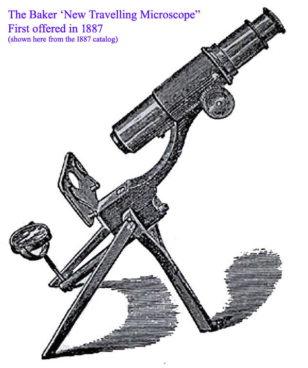 The Baker 'New Traveling Microscope' was apparently first offered by Charles Baker in the 1887 catalog(left), and is the instrument pictured on this page above. It is the successor to the Baker Moginie Type of 'Traveling' Portable microscope, and the precursor of the 'Diagnostic' Model (see below). The usual form of the New Traveling model had only coarse focus by rack and pinion, no fine focus and no condenser. It was supplied for £2 5 with an eyepiece but no objective. However, it could be supplied with only push-pull coarse focusing for only £1 15, and it was also noted that a 'Substage tube can be fitted to carry all apparatus.' This was evidently a brass sleeve that could be screwed into the hole in the stage from the bottom, as this hole is threaded. From 1887 to 1895, the catalog engraving and description of the 'New Portable Traveling' Microscope remained unchanged. In 1896, it was no longer 'New' but simply the 'Portable Traveling Microscope.' In 1899 it was no longer listed in the catalog, apparently replaced entiredly by the various iterations of the 'Diagnostic' model(see below).
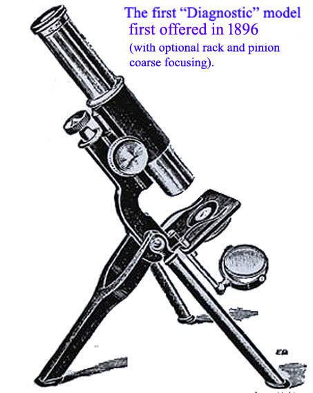
 Sir Ronald Ross, upon discovering that a similar, if not identical microscope, supplied to him by Baker could not easily visualize malarial parasites, suggested to Baker they construct a very similar instrument but with superior capabilities, to be used in the field to identify malarial parasites. The Baker 'Diagnostic Model', first reported in the JRMS of 1896, and first shown in the Baker catalog of 1896(left), was the result. Instead of a rack and pinion coarse focus, the first early models had a push-pull coarse focus but had the addition of two features critical for visualizing things as small as malarial parasites. These were, firstly a fine focus mechanism and secondly, a substage condenser. Another feature of note is that, although the the two front folding legs of the 'New Portable Traveling Microscope' were tapering rectangles, all the 'Diagnostic' models had front legs with a round profile. The base price was £3 10 0 with an eyepiece and the condenser. Rack and pinion coarse focusing was an option and in addition, an iris diaphragm was offered for an additional £0 12 6. A 1/12 oil immersion objective cost another £5 however-more than the entire microscope. Coarse focus was less important when studying only blood smears on slides of consistent thickness, but fine focus was critical as was a good light source. Another reason Ross may have not requested the rack and pinion coarse focus is that in the tropics, the mechanism would become quite loose as the grease liquified, where as the push-pull mechanism would not be liable to such a problem. As time passed, the Diagnostic model was offered with additional features & options(see below). In the JRMS of 1902, it was announced that the Baker 'Portable Diagnostic Microscope' was then made of 'magnalium' an alloy of aluminum and manganese, further reducing its weight. This form was apparently not made for very long, as by 1909, the Baker catalog notes it was again made of brass. I would imagine that this lightweight alloy was too susceptible to damage for rugged portable use in the field.
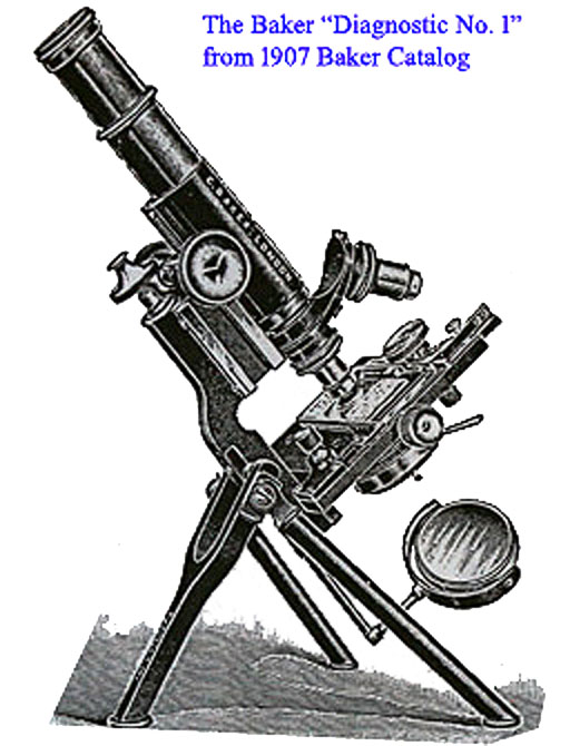
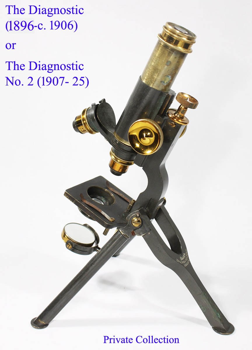
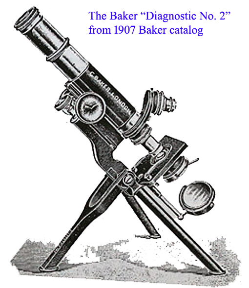 Rack and pinion coarse focus was an option for the Diagnostic model no later than 1899. By 1907, 'diagonal' rack and pinion was standard for coarse focusing, and there were two models of the Diagnostic, the 'Diagnostic No.1(right) and the 'Diagnostic No. 2(left). As you can see, the 'Diagnostic No. 2' was the same as the 'Diagnostic' from previous years. The No. 1 had a folding stage whereas the No. 2 had a fixed stage. Furthermore, a mechanical stage was usually supplied with the No. 1. In addition, the shape of the fine focus knob on the No. 1. was different than the other models. 'Screw focusing' to the substage with centering screws was also an option from 1907 until the 1920's
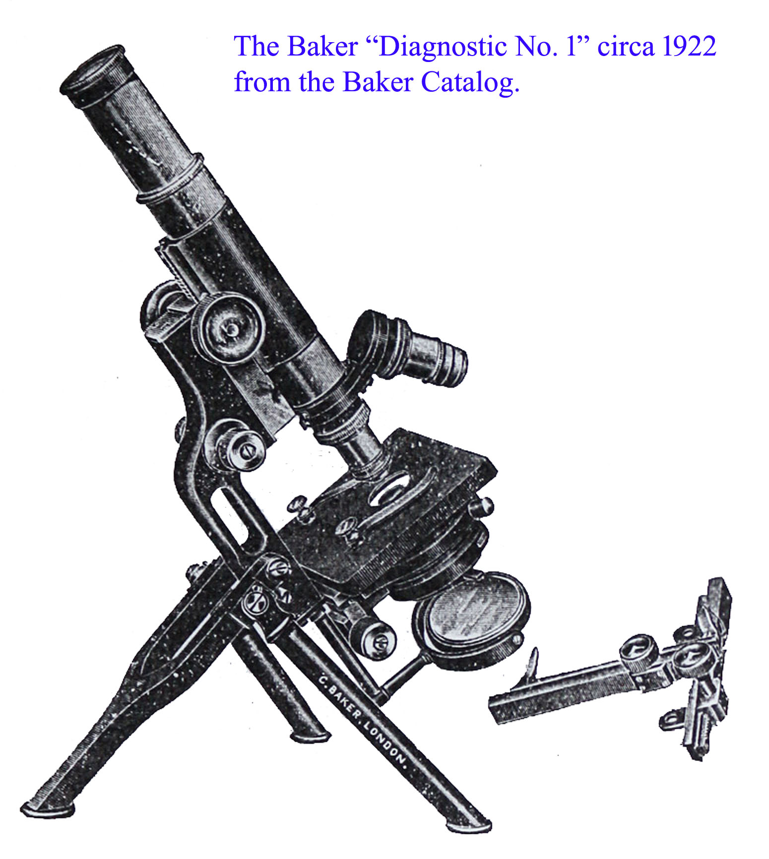
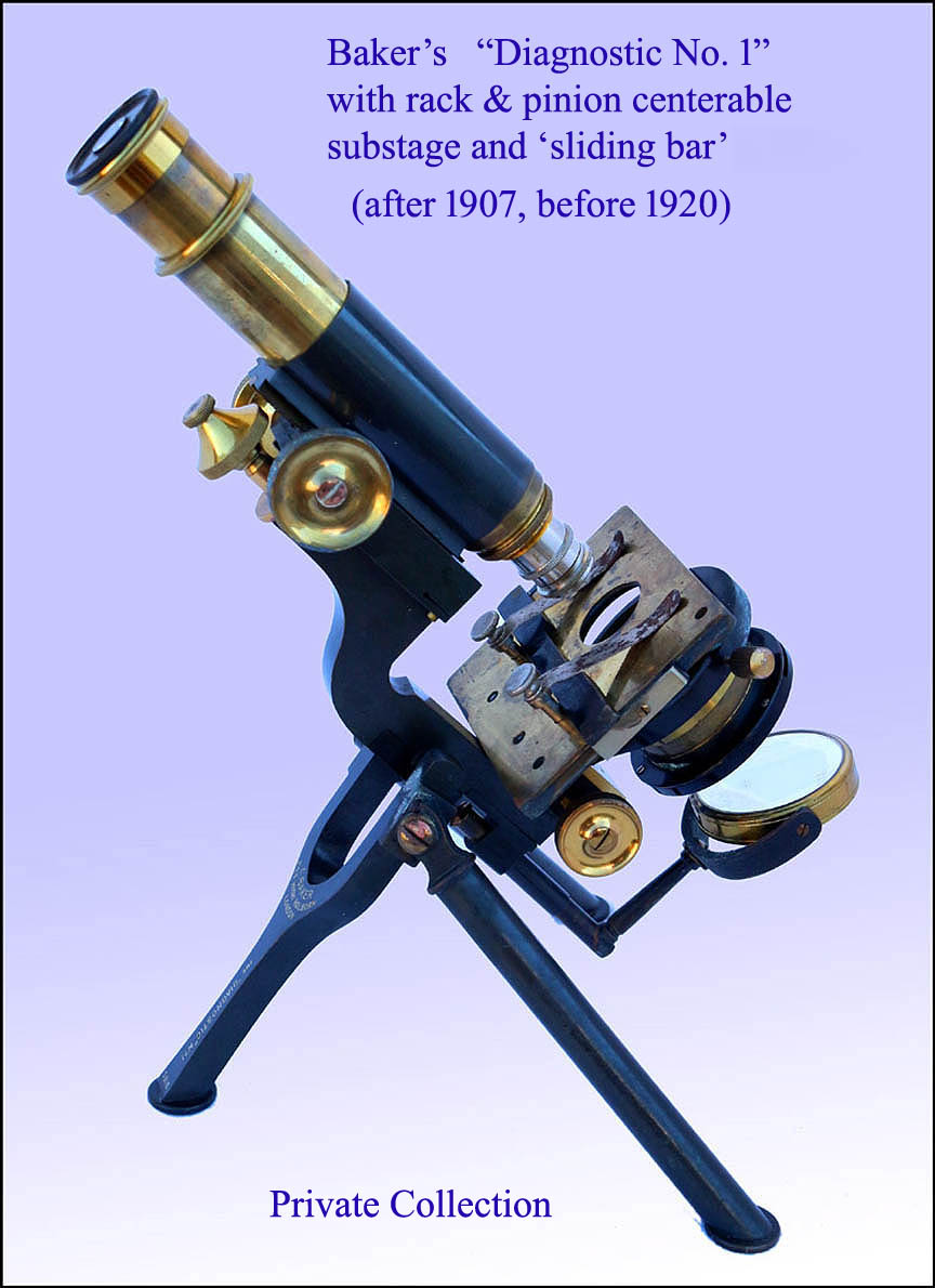 Starting sometime after 1911 but by about 1920, another improved option was introduced: rack and pinion adjustment to the substage. This is shown in the example of the Baker Diagnostic No 1 to the left from sometime after 1911. That microscope shows the substage rack and pinion focusing knob, and it also has a 'sliding bar' for added slide support on the stage. The stand is now more robust allowing a 'more efficient' mechanical stage. Lastly, the substage sleeve is larger, now accepting standard substage accessories. In the slightly later model shown to the right, from the early 1920's, the side fine adjustment was introduced and shown in the engraving as well. This version of the Baker Diagnostic No. 1 continued to be produced through the 1920's, as was the smaller Diagnostic No 2. The No. 2 of the 1920's could be fitted with an Abbe condenser with iris diaphragm, and also with a mechanical stage which was smaller than the one used for the larger No. 1. Diagnostic. The successor to this model was a diagnostic model in a mostly black and chrome finish as seen below.
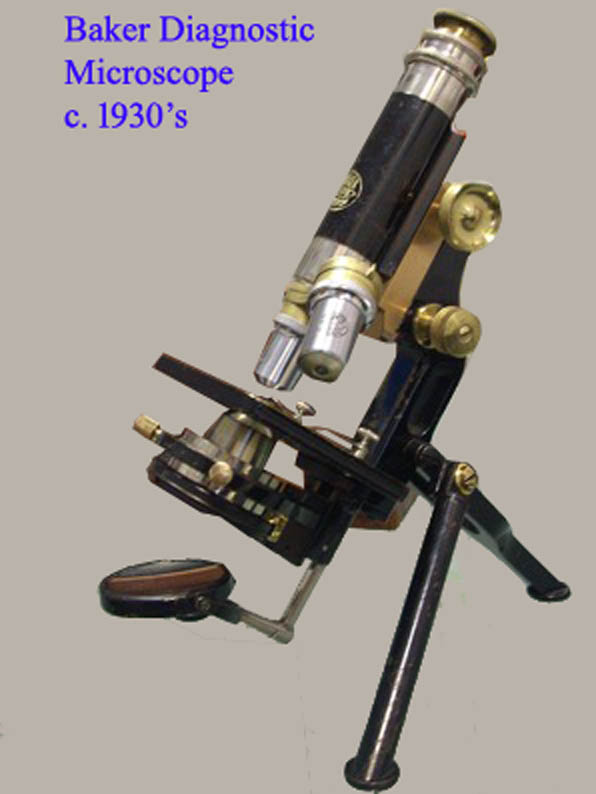 The microscope shown here may be the final form of the Baker Diagnostic, and likely dates to the 1930's. Note the chrome plated tube.